Actinomycetes
| Home | | Pharmaceutical Microbiology | | Pharmaceutical Microbiology |Chapter: Pharmaceutical Microbiology : Characterization, Classification and Taxonomy of Microbes
The Actinomycetes [s., actinomycete], according to the latest edition of Bergey’s Manual (Volume 4), represent an aerobic, Gram-positive bacteria which predominantly and essentially give rise to specific branching filaments or asexual spores or hyphae.
Actinomycetes
The Actinomycetes [s., actinomycete], according to the latest edition of Bergey’s Manual (Volume 4), represent an aerobic, Gram-positive bacteria which
predominantly and essentially give rise to specific branching filaments or asexual
spores or hyphae. It has
been duly observed that the elaborated morphology, arrangement of spores,
explicit cell-wall chemistry, and above all the various kinds of carbohydrates
critically present in the cell extracts are specifically vital and equally
important requirement for the exhaustive taxonomy of the actinomycetes. Consequently, these informations are utilized
meticulously to carry out the articulated division of these bacteria into
different well-defined categories with great ease and fervour. It is quite
pertinent to state at this juncture, that the actinomycetes do possess and exert an appreciable practical impact by virtue of the fact that they
invariably play an apparent major role
in the following two highly
specialized and particular aspects, namely:
(a) Mineralization of organic matter in the
soil, and
(b) Primary
source of most naturally synthesized
antibiotics.
General Characteristics
The
general characteristics of the actinomycetes
are as stated under :
(a) The
branching network of hyphae usually
developed by the actinomycetes,
grows critically both on the surface of the solid
substratum (e.g., agar) as well
as into it to give rise to the formation of substrate mycelium. However, the septate**** mostly divide the hyphae
into specific elongated cells (viz.,
20 μm and even longer) essentially
consisting of a plethora of nucleoids*****.
(b) Invariably,
the actinomycetes afford the
development of thallus. Noticeably,
a large cross-section of the actinomycetes
do possess an aerial mycelium that
extends above the solid subtratum, and produces articulately asexual, thin-walled
spores known as conidia [s., conidium] or conidiospores at the terminal ends of
filaments. In an event, when the spores are located strategically in a
sporangium, they are termed as sporangiospores.
(c) The
spores present in the actinomycetes not only vary widely in terms of shape and
size, but also develop them (spores) by the help of septal formation at the
tips of the filaments, invariably in response to nutrient deprivation. Besides,
a larger segment of these spores are specifically devoid of any thermal
resistance; however, they do withstand dessication quite satisfactorily, and
thus exhibit considerable adaptive value.
(d) Generally,
most actinomycetes are not found to be motile,* and the motility is
particularly confined to the flagellated spores exclusively.
In the
recent past, several taxonomically characteristic features and useful
techniques are of immense value and worth, such as:
·
Morphological features and the colour of mycelia and sporangia
·
Surface properties and arrangement of conidiospores
·
% (G + C) in DNA
·
Phospholipid
content and composition of cell membranes
·
Thermal
resistance encountered in spores
·
Comparison of 16S
rRNA sequences and their values
·
Production of relatively larger DNA fragments by means of restriction enzyme digestion, and
·
Ultimate separation and comparison of ‘larger DNA fragments’ by the aid of Pulsed Field Electrophoresis.
Significance of Actinomycetes
There
are, in actual practice, three most
important practical significances of
the actinomycetes, as mentioned
below:
(1) Actinomycetes are
predominantly the inhabitants of soil and are distributed widely.
(2) They
are able to degrade a large variety and an enormous quantum of organic chemical
entities. However, these are of immense significance in the mineralization of
organic matter.
(3) They
invariably and critically give rise to a large excess of extremely vital ‘natural antibi-otics’ that are used
extensively in the therapeutic armamentarium e.g., actinomycetin. Im-portantly, a
plethora of actinomycetes represent
free-living microbes, whereas a few are pathogens to human beings, animals, and
even certain plants.
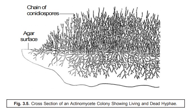
Fig. 3.5.
illustrates the cross-section of an actinomycete colony with living and dead hyphae.
The substrate and aerial mycelium having chains
of conidiospores have been depicted evidently.
Classification
The actinomycetes have been duly classified
into three major divisions based upon
the follow-ing characteristic features:
(a) Whole
cell carbohydrate patterns of aerobic actinomycetes
(b) Major
constituents of cell wall types of actinomycetes,
and
(c) Groups
of actinomycetes based on whole cell
carbohydrate pattern and cell wall type.
The
aforesaid three major divisions shall
now be dealt with separately in the sections that follows.
(a) Whole Cell Carbohydrate Patterns
of Aerobic Actinomycetes
The aerobic actinomycetes do have four distinct whole cell carbohydrate
patterns as given in the following Table 3.5.
Table 3.5. Whole Cell Carbohydrate Patterns of
Aerobic Actinomycetes
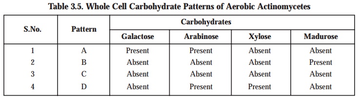
The above
contents of Table: 3.5 vividly shows that none of the four carbohydrates are present in the Pattern ‘C’, whereas Pattern
‘B’ contains only madurose, and Pattern ‘A’ and Pattern ‘D’ con-tains each two
carbohydrates out of the four cited above.
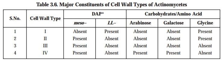
(b) Major Constituents of Cell Wall Types of Actinomycetes
The actinomycetes that possess major
constituents of cell wall types also exhibit four different varieties as provided in Table 3.6.
(c) Groups of Actinomycetes Based on
Whole Cell Carbohydrate Pattern and Cell Wall Type
There are
in all five different varieties of cell wall types having carbohydrate and genera vari-ants in groups of
actinomycetes, as given in Table 3.7 under :
Table 3.7. Actinomycetes Based on Whole Cell
Carbohydrate Pattern and Cell Wall Type
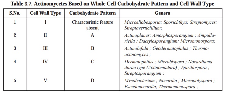
One may
observe from Table 3.7 that the cell
wall type I is devoid of the characteristic feature pertaining to the
specific carbohydrate pattern.
Actinomycetes with Multiocular**
Sporangia***
The
latest version of Bergey’s Manual
has explicitly described the actinomycetes
occurring as the ‘clusters of spores’
in a specific situation when a hypha
undergoes division both transversely
and logitudinally. In reality, all
the three genera critically present
in this section essentially possess chemotype III cell walls, whereas the cell
extract carbohydrate patterns differ prominently.
Salient Features: The
salient features of the actinomycetes with multiocular sporangia are as follows :
(1) The
mole % (G + C) values varies from 57 to 75.
(2) Chemotype III C Cell Walls****: Geodermatophillus belonging to this
category has motile spores and is
specifically an aerobic soil organism.
(3) Chemotype III B Cell Walls : Dermatophillus invariably gives rise to
pockets of motile spores having tufts
of flagella. It is a facultative
anaerobe and also a parasite of mammals actually responsible for the skin
infection streptothricosis.
(4) Chemotype III D Cell Walls: Frankia usually
produces non-motile sporangiospores evi-dently located in a sporogenous body.
It is found to extend its normal growth in a symbiotic association particularly
with the roots of eight distinct families
of higher non-leguminous plant sources viz., alder trees. These
organisms are observed to be extremely efficient microaerophilic nitrogen-fixers which frequently take place very much
within the root nodules of the plants. Furthermore, the
roots of the infected plants usually develop nodules that would eventually cause fixation of nitrogen so efficiently
that a plant, for instance : an alder
tree, may grow quite effectively even in the absence of combined N2,
when nodulated respectively. It has
been duly observed that very much inside the nodule cells, Frankia in-variably gives rise to branching hyphae having globular vesicles strategically located
at their ends. Consequently, these vesicles could be the most preferred sites
of the N2 fixation ultimately. However, the entire phenomenon of N2
fixation is quite similar to that of Rhizobium
wherein it is both O2 sensitive and essentially and predominantly
needs two elements, namely : molybdenum
(Mo), and cobalt (Co).
Related Topics
