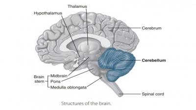
Brain Protection
| Home | | Anatomy and Physiology | | Anatomy and Physiology Health Education (APHE) |Chapter: Anatomy and Physiology for Health Professionals: Central Nervous System
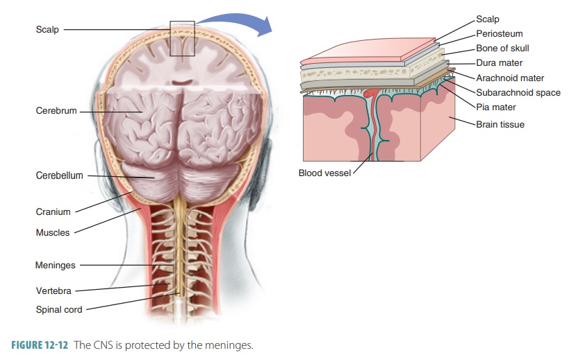
The brain is surrounded by bones, fluids, and membranes. It lies inside the skull’s cranial cavity and is soft and delicate.
Brain
Protection
The brain is surrounded by bones,
fluids, and membranes. It lies inside the skull’s cranial cavity and is soft
and delicate. Between the bony coverings and the soft brain tissues are layered
membranes known as meninges that
protect the brain and spinal cord (FIGURE 12- 12). The singular term meninx
describes just one of the meninges. Also, the CSF cushions the brain and the blood–brain barrier protects it from
harmful substances carried in the blood. There is also a blood–CSF barrier, which is formed by specialized ependymal cells
surrounding the capillaries of the choroid plexus.

Meninges
There are three layers of
membranes in the menin-ges: the dura mater, arachnoid mater, and pia mater. The outermost layer (dura mater) is made up of fibrous, tough,
white connective tissue. It has many blood vessels and nerves and attaches to
the inside of the cranial cavity; it also extends inward between the brain
lobes to form protective partitions. It con-tinues into the vertebral canal to
surround the spinal cord, ending in a sac at its end. The dura mater has two
layers of fibrous connective tissue. Its periosteal
layer, which is more superficial,
attaches to the peri-osteum (the inner surface of the skull). Around the spinal
cord, there is no dural periosteal layer. The meningeal layer actually covers the brain, continu-ing caudally as
the spinal dura mater in the vertebral canal. The two dural layers of the brain
are fused in most areas. In certain places they separate, enclosing dural venous sinuses , which collect blood from
the brain and channel it to the
internal jugular veins in the neck.
Dural septa limit excessive brain movement and are formed from the
meningeal dura mater. It con-tains three primary features. Its falx cerebri is a large fold that dips into the longitudinal fissure between
the hemispheres of the cerebrum. It contains two large venous sinuses: the superior sagittal and the inferior sagittal. The falx cerebelli continues inferiorly from the
more posterior falx cerebri along the vermis of the cerebellum. The tentorium cerebelli is a nearly hor-izontal dural fold extending into the transverse
fissure and cerebellum. It contains the transverse
sinus.
The membrane around the spinal cord has an epidural space separating it from the vertebrae. The epidural space contains loose adipose and con-nective tissues, protecting the spinal cord. The thin, web-like arachnoid mater lies between the dura and pia maters. The subdural space is a narrow serous cavity that contains a fluid film.
The thin pia mater has many blood
vessels and nerves that nourish the brain and the spinal cord. The pia mater is
closely aligned with the surfaces of these organs. It is comprised of many tiny
blood vessels and delicate connective tissue. The pia mater is bound tightly to
the brain and its convolutions. Small ragged bits of pia mater are briefly
carried by small arteries entering the brain. There are also spinal meninges, discussed later.
Cerebrospinal Fluid
Between the arachnoid and pia
maters is a sub-arachnoid space containing the watery and clear cerebrospinal fluid (CSF). This is similar to blood plasma
but contains less protein. CSF contains more sodium, chloride, and hydrogen
ions but less calcium and potassium than blood plasma. Inside the sub-arachnoid
space are web -like extensions that func-tion partially to bind the arachnoid
mater to the pia mater. This space also contains CSF and the primary brain
blood vessels. However, these blood vessels are not protected well because the
arachnoid mater is very fine and elastic. Arachnoid
villi are knob-like projections that
protrude superiorly through the dura mater into the superior sagittal sinus.
They absorb CSF into the venous blood of the sinus. In adults, clusters of
arachnoid villi form large arachnoid granulations, where CSF is actually absorbed into the venous circulation.
Small red choroid plexuses secrete
CSF and project into the brain ventricles. Most CSF is formed in the lateral
ventricles. CSF also enters the menin-ges’ subarachnoid space via the two lateral apertures and the single median aperture and is reabsorbed into
the blood. CSF surrounds the brain and spinal cord, maintaining a stable ionic
concentration and protect-ing CNS structures. The brain floats in CSF, which
cushions it and prevents the bottom of the brain from being crushed by its own
weight. The CSF also helps to nourish the brain and may assist in carrying chemical
signals concerning sleep and appetite. It also may carry hormones. The total
CSF volume is about 150 mL, which is replaced every eight hours.
Blood Supply to the Brain
The human brain’s billions of
neurons continuously demands oxygen and nutrients. The neurons do not have
carbohydrates or lipids as energy reserves. They also do not have myoglobin as
an oxygen reserve. Therefore, the blood supply to the brain is extensive.
Arterial blood is supplied through the internal
carotid arteries and vertebral arteries. The majority of the brain’s venous blood leaves the cranium
through the internal jugular veins that
drain the dural sinuses. If a head
injury damages cerebral blood vessels, there may be bleeding into the dura
mater. This can occur near the dural epithelium or between the outer dura mater
and skull bones. Both are serious conditions, since blood entering these spaces
will compress and distort the brain’s soft tissues.
Blood–Brain Barrier
The brain requires a constant
internal environment to function normally. To maintain this, the blood– brain
barrier acts to selectively
allow certain mol-ecules to pass and to keep others from reaching the brain.
The maintenance of a constant environment keeps the brain’s neurons from firing
uncontrolla-bly. Before bloodborne substances can move from brain capillaries
to reach neurons, three layers of the blood–brain barrier await them: the
capillary wall endothelium, a thick basal lamina surround-ing every capillary’s
external side, and bulb-like feet of
the astrocytes that are bound to the capillaries.
These portions of astrocytes give
signals to the endo-thelial cells and cause them to form tight junctions. The junctions form the actual barrier by causing a
seamless pattern of endothelial cells. This capillary barrier is the least
permeable of all capillary struc-tures in the body.
The selective blood–brain barrier
allows certain electrolytes, nutrients, and essential amino acids to pass
through. It does not allow passage of proteins, bloodborne metabolic wastes,
most drugs, and specific toxins. Also, nonessential amino acids and potassium
ions are actively pumped across the capillary endothe-lium away from the brain.
The blood–brain barrier is not effective against fatty acids, fats, carbon
dioxide, oxygen, and other fat-soluble molecules that can easily diffuse
through the body’s plasma membranes. There-fore, bloodborne anesthetics,
alcohol, and nicotine affect the brain.
The blood–brain barrier is not
uniform through-out the brain and is even absent near the third and fourth
ventricles. In the brain’s vomiting center, blood-borne molecules can easily
cross to the neural tissue, but this is part of this center’s monitoring for
poisons. The hypothalamus also does not have a barrier, allow-ing it to sample
the blood’s chemical composition so it can regulate many metabolic activities.
In newborns and premature infants, the blood–brain barrier is not fully formed.
As a result, certain toxins can enter the CNS and cause certain conditions that
do not gener-ally affect adults. Also, the blood–brain barrier can be broken
down by brain injuries. When these occur, the capillary endothelial cells or
their tight junctions are commonly affected.