Glands in the Skin
| Home | | Anatomy and Physiology | | Anatomy and Physiology Health Education (APHE) |Chapter: Anatomy and Physiology for Health Professionals: Support and Movement: Integumentary System
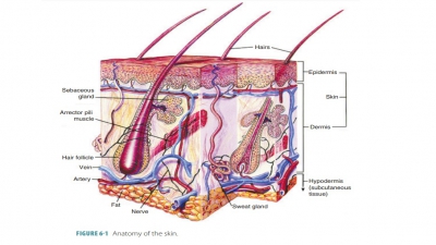
The skin contains two types of exocrine glands: sebaceous glands and sweat glands.
Glands in the
Skin
The skin contains two types of
exocrine glands: sebaceous glands and sweat glands. The sebaceous (oil) glands
are simple and branched alveolar glands cov-ering the body, except on the palms
and soles. The sweat (sudoriferous) glands are found all over the body except
for the lips, nipples, and certain parts of the external genitalia.
Sebaceous Glands
Sebaceous
glands (oil glands) are made
up of spe-cialized epidermal cells and are primarily located near hair
follicles. These glands are largest on the face, neck, and upper chest. They
are actually holocrine glands, secreting sebum , which is an oily mixture of fatty material and debris
from cells. The central alve-oli cells accumulate lipids until they burst, and
the combined lipids and cell fragments make up sebum. The sebum is secreted
through small hair follicle ducts, helping to keep both hair and skin pliable
and waterproof. The sebum is a mixture of cholesterol, triacylglycerides,
proteins, and electrolytes. Sebum inhibits bacterial growth, protecting the
keratin of the hair shafts. Sebum is forced out of hair follicles to the skin
surface via arrector pili contractions. This lubricates the hair and skin, keeping
the hair supple and slowing the loss of water from the skin during times of low
environmental humidity. Sebum has a strong bactericidal action. Its secretion
is stimulated by androgens, primarily. Hence, sebaceous glands are less active
until a human reaches puberty and andro-gen production rises.
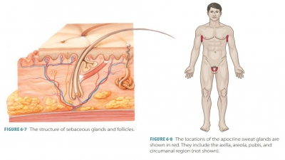
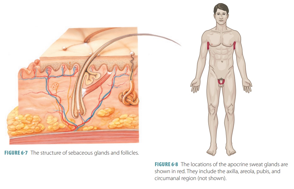
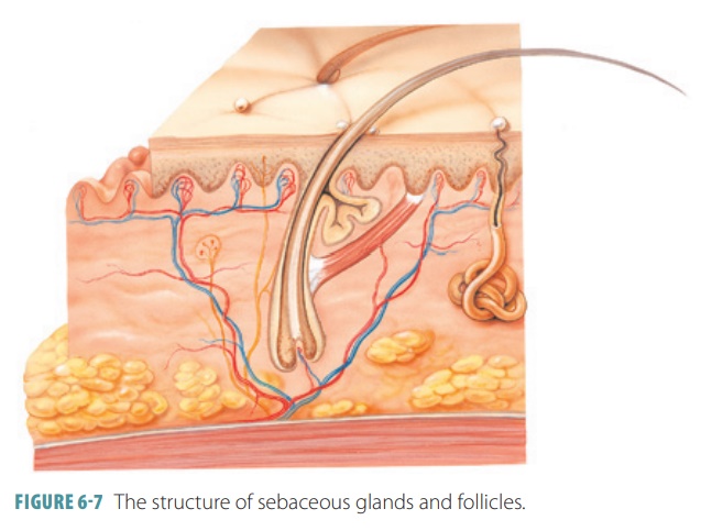
Sebaceous
follicles are large sebaceous
glands that surround hair follicles. Their ducts discharge sebum
directly onto the epidermis (FIGURE 6-7). They are found on the face, chest, nipples, back, and
exter-nal genitalia. During the final phases of fetal develop-ment, their
secretions as well as epidermal cells that have been shed coat the skin surface
to form a protec-tive layer. When the sebaceous glands become overac-tive,
usually occurring on the scalp, an inflammation may develop around them. This
is known asseborrheic dermatitis, which is a common cause of
dandruff.
Sweat Glands
Sweat glands consist of a small
tube originating as a coil in the deep dermis or superficial subcutaneous
layers. The coiled portion is lined with sweat-secreting epithelial cells.
Sweat is carried out of the skin by tubes called pores that open at the skin
surface. Sweat is made up of 99% water as well as salts, which are primarily
sodium chloride, ascorbic acid, or vitamin C; antibodies; and waste products,
including urea, ammonia, and uric acid, sweat tastes salty because of its
electrolytes. Sweat also contains dermicidin, which is a peptide that kills microbes. Overall, sweat is a hypotonic
filtrate of blood, passing through secre-tory cells via exocytosis. Its
composition is based on diet, heredity, and partially certain drugs that are
ingested. Sweat has a normal acidic pH of between 4 and 6. Sweating is
regulated by the autonomic ner-vous system to prevent overheating. It begins on
the forehead, spreading inferiorly to the rest of the body. When sweating is
brought about by nervousness or fright (cold sweating), it starts on the palms,
axillae, and soles before spreading throughout the body.
The skin contains two types of sweat glands (sudoriferous glands): merocrine sweat glands and
apocrine sweat glands. Merocrine
(eccrine) glands are the predominant type of sweat glands, responding to body
temperature, and are present at birth. They excrete water and electrolytes and
also provide protec-tion from hazards in the environment. Adult skin con-tains
two to five million merocrine sweat glands. They are found on the forehead,
neck, and back, although the palms and soles have the highest numbers. They are
simple tubular glands with a coiled appearance. The secretory portion is found
in the dermis, whereas the duct opens in a funnel-shaped pore at the surface of the skin. These pores are not the same as
the “com-plexion pores,” which are the outlets of hair follicles.
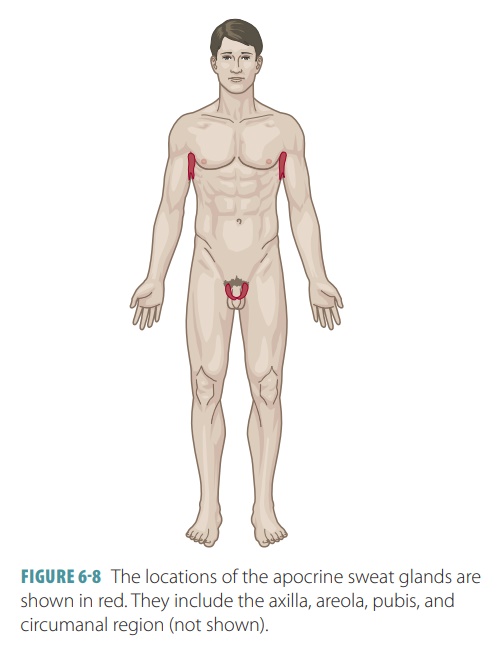
Apocrine glands are sweat glands
that become active at puberty and number about 2,000. They are found mostly in
the armpits and groin, with the sweat excreted at these places developing a
scent as they come into contact with skin bacteria (FIGURE
6-8). This is the basis of body
odor. Modified sweat glands include the ceruminous
glands of the external ear
(which pro-duce earwax) and the mammary glands (which produce milk). Cerumen or earwax is believed to block
entry of foreign materials or insects into the ear.
It should be noted that apocrine
glands are still actually merocrine glands that produce their product in the
same way as eccrine sweat glands. However, they are larger in size, are located
in the dermis or hypo-dermis, and empty into hair follicles. Their secretions
are not only similar to eccrine glands, but also include proteins and fatty
substances. The color of these secretions may be white or yellow. The function
of apocrine glands is controlled by androgens, activated by sympathetic nerve
fibers during stress and pain. In women, they enlarge and recede along with the
men-strual cycle. The secretory cells of apocrine glands are surrounded by myoepithelial cells that
squeeze them to discharge accumulated sweat into the hair follicles.
1. Describe
how hairs grow out of the skin.
2. Distinguish
between eccrine and apocrine glands.
3. What is
the function of the sebaceous glands?