Methods for Studying Drug Uptake
| Home | | Biopharmaceutics and Pharmacokinetics |Chapter: Biopharmaceutics and Pharmacokinetics : Absorption of Drugs
The experimental methods for studying absorption of drugs can be classified as in vitro and in situ.
METHODS FOR STUDYING DRUG UPTAKE
The experimental methods for studying absorption of drugs can be classified as in vitro and in situ.
1. In vitro experiments: are used to study the transport of drugs through different types of membranes or biological materials. Such experiments may utilize
(a) Diffusion cells
(b) Segments of GIT of laboratory animals – Two well known established techniques are –
(i) Everted sac technique, and
(ii) Everted ring technique.
(c) Cell cultures of gut epithelium e.g. Caco-2 cells.
2. In situ experiments: simulates the in vivo conditions for drug absorption and are based on perfusion of a segment of GIT by drug solution and determination of amount of drug diffused through it. The two perfusion methods used in laboratory animals are –
(a) Doluisio method
(b) Single pass perfusion.
1a. Diffusion Cell Method
Diffusion cells consist of two compartments –
1. Donor compartment which contains the drug solution and the lower end of which contains the synthetic or natural GI membrane that interfaces with the receptor compartment.
2. Receptor compartment which contains the buffer solution.
The procedure of uptake study using this technique involves measurement of rate of arrival of drug in the receptor compartment (see figure 2.32).
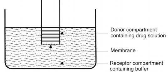
Fig. 2.32. Diffusion cell for studying drug uptake from GIT.
1b. (i) Everted Sac Technique
This technique involves eversion of a segment (about 3 cm) of the intestine of small intestine, conversion into a sac filled with buffer solution and its immersion into a bath containing oxygenated solution of drug. The whole preparation is maintained at 370C and shaken mildly. A predetermined time intervals, the sac is removed and the concentration of drug in the serosal (internal) liquid is determined (see figure 2.33).
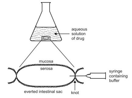
Fig. 2.33. Everted sac technique for studying drug transport. uptake from GIT.
1b.(ii) Everted Ring Technique
In this technique, appropriate section of intestine of rat is isolated, inverted and dissected in slices or rings of thickness between 1 and 3 mm. The slices are then placed in an oxygenated isotonic drug solution at 370C and incubated for a predetermined period under gentle shaking. After incubation, the rings are washed, dried and placed in a pre-weighed scintillation vials. Each vial is reweighed to determine the wet tissue weight, and then the sample is analysed for drug.
1c. Cell Culture Technique
In this technique, differentiated cells of the intestine, originating from Caco-2 cells (cells of carcinoma of colon) are placed on synthetic polycarbonate membrane previously treated with an appropriate material such as collagen which on incubation aids reproduction of cells while not retarding drug permeation characteristics. Solution of drug is placed on this layer of cultured cells and the system is placed in a bath (receptor compartment) of buffer solution. The drug that reaches the latter compartment is sampled and analysed periodically.
2a. Doluisio Method
In this method, the upper and lower parts of the small intestine of anaesthetised and dissected rat are connected by means of tubing to syringes of capacity 10 – 30 ml (see figure 2.34). After washing the intestinal segment with normal saline, the syringe is filled with a solution of radiolabelled drug and a non-absorbable marker which is used as an indicator of water flux during perfusion. Part of the content of the syringe containing drug is delivered to the intestinal segment which is then collected in the second syringe and analysed for drug.
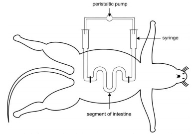
Fig. 2.34. Doluisio method for drug uptake through rat intestine.
2b. Single-Pass Perfusion
In this technique the drug solution passes through the intestinal segment just once (see figure 2.35). This technique is superior to Doluisio method in that precise adjustment of hydrodynamic conditions that can influence blood circulation and puts stress on intestinal wall can be controlled.
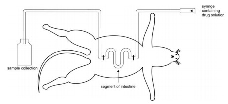
Fig. 2.35. Single-pass perfusion technique for studying drug uptake.
Related Topics
