Mouth
| Home | | Anatomy and Physiology | | Anatomy and Physiology Health Education (APHE) |Chapter: Anatomy and Physiology for Health Professionals: Digestive System
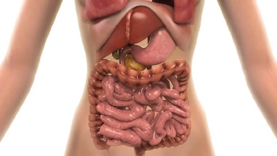
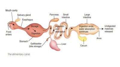
Digestion begins in the mouth, which is also known as the oral cavity or buccal cavity.
Mouth
Digestion begins in the mouth,
which is also known as the oral
cavity or buccal cavity.
It is encompassed superiorly by the palate, anteriorly by the lips, laterally
by the cheeks, and inferiorly by the tongue. The oral orifice consists
of the mouth’s anterior opening. The mouth
is continuous posteriorly with the oropharynx.
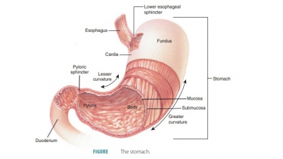
Solid particles of food are reduced mechanically and mixed with saliva. The vestibule is the portion that
lies between the teeth, cheeks, and lips.
Tongue
The tongue is
covered by a mucous membrane and is connected to the floor of the mouth by a
membranous fold called the lingual
frenulum. The body of the tongue is made up of primarily interlaced,
bundled skel-etal muscle. It mixes food particles with saliva during chewing,
moving food, in the form of a mass called a bolus, toward the pharynx during swallowing. The tongue also moves food underneath the teeth for
chew-ing and helps to form consonant sounds when speaking.
The tongue contains intrinsic and extrinsic skele-tal muscle
fibers. Its intrinsic
muscles are within the tongue itself and not attached to any bones.
However, its extrinsic
muscles originate on either the skull bones or soft palate,
extending to the tongue. They change the tongue’s position and are able to move
it left and right and cause it to protrude or retract. There is a median septum
of connective tissues, with each half containing identical muscle groups.

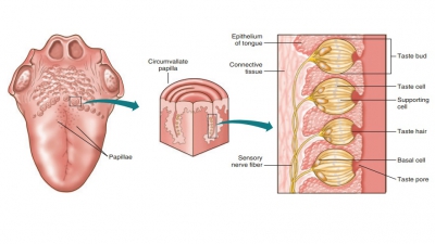
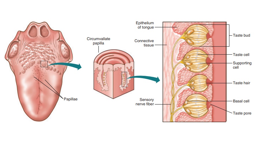
The tongue has rough papillae that project from its surface to provide friction (FIGURE 24-5). These peg-like projections
emerge from the mucosa.The filiform papillae are cone-shaped and make the
tongue’s surface rougher. They aid in eating semisolid foods, providing
friction so these foods can be manip-ulated. They are found in parallel rows on
the dorsum of the tongue and are the smallest, most numerous type of papillae.
Keratin is contained within the fili-form papillae, causing the tongue to have
a somewhat white appearance. The keratin makes the filiform papillae stiff in
comparison with other papillae.
Over much of the tongue’s surface are scattered fungiform papillae, which are mushroom
shaped. They
have a red appearance because of their vascular cores. In a V-shaped row on the
posterior tongue are 10–12 vallate
papillae. These papillae are very large, appear similar
to the fungiform papillae, and have a furrow that surrounds them. On the
lateral aspects of the posterior tongue are found foliate papillae, which appear as if they
are folded into “pleats.” The fungiform, vallate, and foliate papillae also
contain the taste buds. In infancy and early childhood, the taste buds of the
foliate papillae are most functional, with reduced function later in life.
There are no papillae on the mucosa that covers the root of the tongue.
Tonsils and Palate
The root of the tongue is connected to the hyoid bone and
covered with rounded lymphatic tissue masses called lingual tonsils, giving it a bumpy texture. The
lingual tonsils lie just deep to the mucosa. The roof of the oral cavity is
formed by the palate, which
consists of a bony, anterior hard palate and a muscular, posterior soft palate.
The soft palate arches posteriorly and down-ward into a cone-shaped projection
called the uvula. In the back
of the mouth, on either side of the tongue and near the palate, are masses of
lymphatic tissue called the palatine
tonsils. They lie beneath the epithelial lining of the mouth and help to protect against infection.
The pharyngeal
arches lie on either side of the uvula, and the more anterior palatoglossal arch is located between the soft palate
and the base of the tongue. The fauces
is the arched opening between the soft palate and base of the tongue and is
formed by a curved line connecting the palatoglossal arches and uvula. The
fauces create the passage between the oral cavity and oropharynx. Extending
from the soft palate to the pharyngeal wall is the more posterior palatopharyngeal arch. One palatine tonsil lies between the palatoglossal and palatopharyngeal arches on either
side.
The pharyngeal
tonsils are also known as the adenoids.
They lie on the posterior pharynx, above the
border of the soft palate. When the adenoids enlarge to block the passage
between the nasal cavity and pharynx, they may be surgically removed, similar
to the palatine tonsils.
1. Describe the structures that encompass the oral cavity.
2. Explain the various functions of the tongue.
3. Differentiate between the various types of tonsils.
4. Describe foliate and vallate papillae.