Muscle Tissues
| Home | | Anatomy and Physiology | | Anatomy and Physiology Health Education (APHE) |Chapter: Anatomy and Physiology for Health Professionals: Levels of Organization : Tissues
Skeletal Muscle Tissue, Smooth Muscle Tissue, Cardiac Muscle Tissue
Types of Tissues
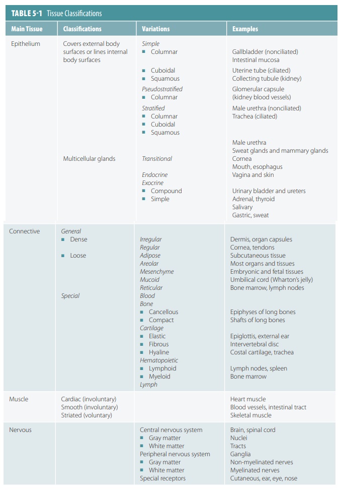
The human body is primarily made
up of four major types of tissues: epithelial, connective, muscle, and nervous.
Epithelial tissues cover body surfaces, cover and line internal organs, and make up the
glands. Connective tissues are widely distributed throughout the body, filling internal spaces,
and func-tion to bind, support, and protect body structures. Muscle tissues are specialized for contraction and
include the skeletal muscles of the
body, the heart, and the muscular walls of hollow organs. Skeletal muscles are
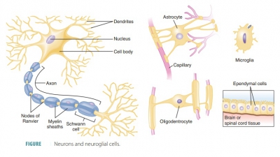
attached to bones and are used for movement of the body. Nervous tissues carry information from one part of the body to another via
electrical impulses. They are found in the brain, spinal cord, and nerves (TABLE 5-1).
The human body is primarily made up of four major types of tissues:
1. epithelial,
2. connective
3. muscle, and
4. nervous.
Muscle
Tissues
Muscle tissues can contract by
shortening their elongated muscle fibers. This action moves body parts. Myofilaments in muscle cells are
responsible for the muscles’ ability
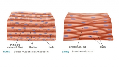
to move or contract. The three types of muscle tissue are skeletal muscle
tissue, smooth mus-cle tissue, and cardiac muscle tissue.
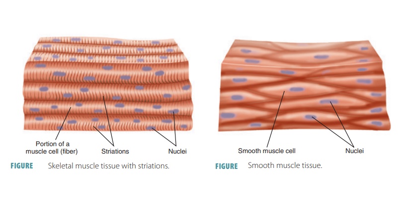
Skeletal Muscle Tissue
Skeletal muscle
tissue is known as voluntary
muscle tissue because it is found in muscles controlled by
con-scious effort. It attaches to bones and is composed of long thread-like
cells that have light and dark markings called striations (FIGURE 5 -9). These multinucleated muscle cells contract when
stimulated by nerve cells. The term “multinucleated” describes the several
hun-dred nuclei distributed inside the muscles’ plasma membranes. Skeletal
muscle tissue moves the head, trunk, and limbs, allowing all voluntary movements
in these body areas. Skeletal muscle cells are also called muscle fibers. They are long and cylindrical, con-taining many peripherally
located nuclei.
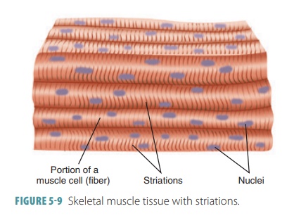
The cells of skeletal muscle tissue
may be one foot or more in length. The fibers cannot divide. New fibers are
produced when satellite cells divide.
These are stem cells that remain in adult skeletal muscle tissue. There-fore,
skeletal muscle tissue can partially repair itself after injury. Skeletal
muscle is also known as striated voluntary muscle. It actually contains
all four types of body tissue, not
just muscle tissue. Adjacent fibers are bound by collagen and elastic fibers
that blend into an attached tension or aponeurosis,
which con-ducts the force of contraction. Movement is, therefore, produced when
skeletal muscles contract and pull on attached bones.
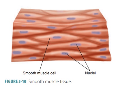
Smooth Muscle Tissue
Smooth muscle
tissue is composed of
elongated, spindle-shaped cells in muscles not under voluntary control.
The cells have tapered ends and a single, oval nucleus. Smooth muscle fibers
are shorter than striated fibers, having only one nucleus per spindle-shaped
fiber. They are also called nonstriated
involuntary muscles or unstriated muscles (FIGURE 5-10). Smooth muscle cells have actin and myosin filaments that are
different from skeletal or cardiac muscle. They may contract on their own. Gap junctions exist between adjacent cells, which
coordinate the contractions of individual cells.
Smooth muscle cells can divide;
therefore, they regenerate after being injured. Smooth muscle tissue composes
hollow internal organ walls (such as the intestines, stomach, urinary bladder,
blood vessels, and uterus). Smooth muscle cannot, in most cases, be controlled
by conscious effort. This type of tissue moves food through the digestive
tract, empties the urinary bladder, and constricts blood vessels. It
accom-plishes these tasks by either contracting or relaxing.
Cardiac Muscle Tissue
Cardiac
muscle tissue is also called the myocardium, the thick middle layer of the heart wall. The contractile
tissue of the myocardium is composed of fibers, with the characteristic,
prominent cross-striations of mus-cular tissue. These striations branch
frequently and are interconnected, forming a network. Myocardial muscle
contains less connective tissue than skeletal muscle and is usually uninucleate
(having one nucleus, located centrally).
However, some cardiocytes have up to five nuclei. Cardiac muscle is
involuntary and makes up most of the heart. It relies on pacemaker cells for
regular contraction.
Cardiac muscle cells, known as cardiocytes, are branched and fit
together tightly at junctions known as intercalated
discs. In these locations, membranes
are joined by desmosomes, gap junctions, and proteo-glycans. As ions move
through the gap junctions, they help synchronize cardiac muscle cell
contractions. The desmosomes and proteoglycans lock the cells together as they
contract. The cells do not rely on nerve activ-ity to begin a contraction;
specialized pacemaker cells establish
regular contraction rates instead. The nervous system has the ability to alter
the rate of pacemaker cell activity. However, it does not provide any
volun-tary control over individual cardiac muscle cells. As a result, cardiac
muscle is called striated
involuntary muscle. Cardiac muscle tissue is not able to signifi-cantly repair
itself. After an injury to the heart, some muscle cells are able to divide.
However, repairs are not complete and some heart function will usually be lost.
1. What
type of muscle cells is striated?
2. Identify
examples of voluntary and involuntary muscles.
3. What
type of muscle cells is branched and has intercalated discs?