Muscular System
| Home | | Anatomy and Physiology | | Anatomy and Physiology Health Education (APHE) |Chapter: Anatomy and Physiology for Health Professionals: Support and Movement: Muscular System
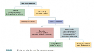
Skeletal muscles usually function in groups, with the nervous system stimulating the desired muscles to perform the intended function.
Muscular
System
After
studying this chapter, readers should be able to:
1. Distinguish
between the origin and insertion of a skeletal muscle.
2. Differentiate
between flexion and extension.
3. Differentiate
between parallel, convergent, pennate, and circular muscles.
4. Which
type of lever system is most common in the skeletal muscles?
5. Identify
the actions of the rhomboid major, subclavius, and trapezius muscles.
6. Explain
the movements of the deltoid, pectoralis major, and teres major.
7. Name
the muscles of the abdominal wall and explain the action of the rectus
abdominis.
8. Explain
the muscles that flex and extend the thigh.
9. Describe
the quadriceps femoris group and the function of the muscles it contains.
10. Describe
the function of the gastrocnemius.
Overview
Skeletal muscles usually function
in groups, with the nervous system stimulating the desired muscles to perform
the intended function. A muscle that con-tracts to provide most of a desired
movement is called a prime mover or agonist. A good example is the pectoralis major muscle, which is a prime mover
of arm flexion. Other muscles, known as synergists, work with a prime mover to make its action more effective by adding a
small amount of additional force. For example, when you bend your forearm, the
ago-nist muscles are the biceps and the synergists are the triceps. In muscles
that cross several joints, contrac-tion causes movement at all the spanned
joints unless other muscles act as stabilizers of the joints. Other flex-ors
may cause some undesirable movements in a joint, but synergists prevent this
and allow the total force of the prime mover to occur in the desired
directions. Some synergists, known as fixators,
may also assist an agonist by preventing another joint from moving to stabilize
the origin of the agonist. Fixator muscles run-ning from the axial skeleton to
the scapula cause the scapula to be immobilized. Only desired movements can
then occur at the shoulder joint. The muscles that help maintain upright
posture are fixators.
Other muscles act as antagonists to prime
mov-ers. They cause movement in the opposite direction. In the earlier example,
the triceps are the antagonists to the biceps. Smooth body movement depends on
antagonists relaxing while prime movers contract. Muscles may work opposite to
each other or together to control various movements. Antagonists and their
prime movers are situated on the opposite sides of joints across which they
function. An example is the pectoralis major, which acts as an antagonist to
the latissimus dorsi, the prime mover that extends the arm. It is important to
understand that antagonists can actually also be prime movers such as
latissimus dorsi when it acts as the prime mover of arm extension.
Origins and Insertions
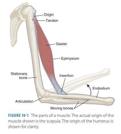
One end of a skeletal muscle
usually is fastened to a relatively immovable part (origin) at a movable joint. The other end connects to a movable part (insertion) on the
other side of the joint. As contraction occurs, the insertion is pulled toward
the origin. There may be more than one origin or insertion such as in the
biceps brachii muscle of the arm. When this muscle contracts, the insertion
being pulled toward its origin causes the forearm to flex at the elbow (FIGURE 10-1). Muscle
con-traction produces specific actions
or movements.
The head of a muscle is the part
closest to its origin. The term flexion
describes a decrease in the angle of a joint, for example, a movement of the
fore-arm that causes it to bend at the elbow. The term extension describes an increase in the angle of a joint, for example, a movement of the
forearm that straightens the elbow.
Arrangement of Skeletal Muscles
Skeletal muscles, according to
the arrangement of their fascicles, are divided into four distinct types:
parallel muscles, convergent muscles, pennate mus-cles, and circular muscles.
The pennate muscles are subdivided into unipennate, bipennate, and multipennate muscles.
Parallel Muscles
Most skeletal muscles are classified as parallel muscles, in which the fascicles are parallel to the long axes. Some are flat muscular bands with broad attachments called aponeuroses at each end, whereas others are thick and cylindrical having tendons at one or both ends. An example of a flat parallel muscle is the sartorius muscle, located in the thigh. When they are thick and cylindrical, they have a spindle shape with a central body. An example of a parallel muscle is the biceps brachii, which has a spindle shape and an expanded body. Sometimes, such spindle-shaped muscles are referred to as fusiform muscles.
Convergent Muscles
In a convergent
muscle, the muscle fascicles extend
over a broad area, converging on a single attachment site. The muscle may pull
on a tendon, aponeurosis, or a slender band of collagen fibers. This band is
known as a raphe. An example of a convergent muscle is the pectoralis major, which is triangular or fan-shaped.
Pennate Muscles
In a pennate
muscle, the fascicles create a com-mon
angle with the tendon and muscle fibers pull at an angle. This means pennate
muscles do not move tendons as far as parallel muscles do, but they have more
tension because they have more muscle fibers and myofibrils. The fascicles and
muscle fibers are short and obliquely attached to the central tendon funning
the entire length of the muscle. An example of a unipennate muscle is the extensor digitorum of the leg, which has
its fascicles inserted into just one side of the tendon. One example of a
bipennate muscle is the rectus femoris in
the thigh, which has fascicles inserted into
the tendon from opposite sides. It resembles the shape of a feather. One
multipennate muscle is the del-toid of
the shoulder, which appears as many feather
located side by side, with each of them inserted into one large tendon.
Circular
Muscles
In a circular
muscle or sphincter, the fascicles are arranged around an opening in a
concentric pattern. Muscle contractions cause a decrease in the diameter of the
opening, such as the orbicularis oris
muscle of the mouth or the orbicularis
oculi muscle of the eye.
Arrangement of Fascicles
The range of motion and strength
of a muscle are based on the arrangement of its fascicles. Skeletal muscle
fibers can shorten to approximately 70% of their resting length when they
contract. Therefore, longer and more parallel muscle fibers, in compar-ison to
a muscle’s long axis, means the more the muscle is able to shorten. Parallel
fascicle arrange-ments offer the most ability to shorten, but are not as strong
as other types of fascicle arrangements. The power of a muscle is based on the
number of muscle fibers in it. More muscle fibers offer more strength. For
example, bipennate and multipennate muscles have more fibers and are very
strong while only shortening slightly.
1. Define
the terms origin and insertion.
2. Differentiate
between flexion and extension.