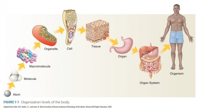
Organization of the Body
| Home | | Anatomy and Physiology | | Anatomy and Physiology Health Education (APHE) |Chapter: Anatomy and Physiology for Health Professionals: Levels of Organization : Introduction to Human Anatomy and Physiology
The human body is composed of distinct body parts, cavities, membranes, and organ systems that include various body systems. All of these are discussed in greater detail in the following sections.
Organization
of the Body
The human body is composed of
distinct body parts, cavities, membranes, and organ systems that include
various body systems. All of these are discussed in greater detail in the
following sections.
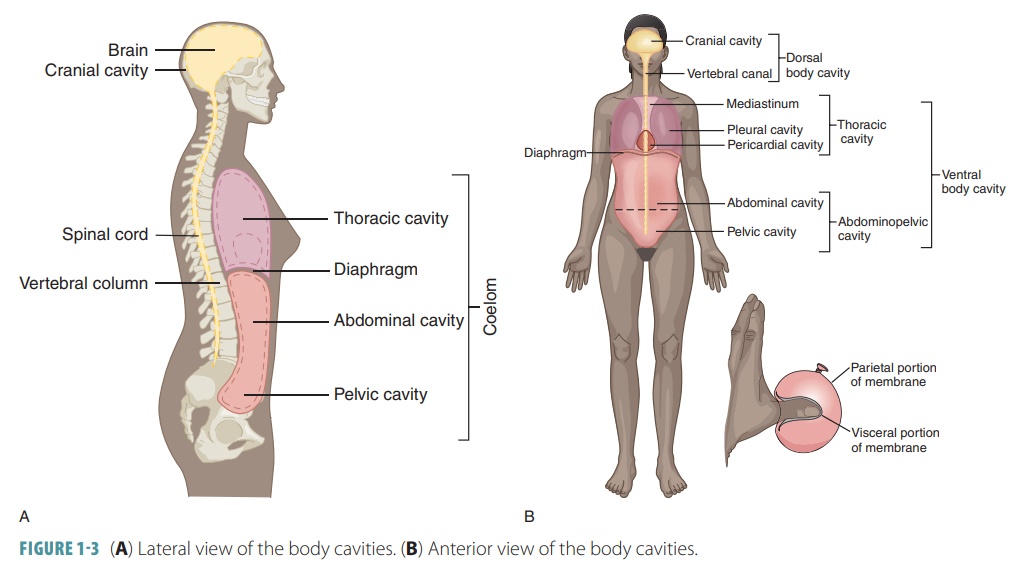
Body Cavities and Membranes
The body is divided into two main
cavities, the dorsal cavity and the ventral cavity. These two main cavities
are divided into smaller subcavities. The dorsal cav-ity protects the organs of
the nervous system. Its two subdivisions include the cranial cavity of the skull, which encases the brain, and the vertebral (spinal)cavity, located inside
the vertebral column, whichencases the spinal cord. The vertebral cavity is
also referred to as the vertebral canal.
The cranial and spinal cavities are in continuation with each other. The
ventral cavity contains most of the body’s organs. More anterior and larger
than the dorsal cavity, it houses the viscera (visceral organs). The ventral cavity is divided into the thoracic cavity and the abdominopelvic cavity.
Thoracic Cavity
The thoracic cavity is surrounded
by the chest mus-cles and ribs, and contains the lungs and heart; organs of the
cardiovascular, respiratory, and lymphatic sys-tems; inferior esophagus; and
the thymus. It is sub-divided into lateral pleural
cavities, which surround each lung, and
the medial mediastinum, which is a tissue mass that separates the cavities. Each pleural
cavity is lined by a serous
membrane, which is shiny and slippery,
and functions to reduce friction as the lung expands and recoils during
breathing. The pleura is the serous
membrane lining a pleural cavity. The visceral
pleuracovers the outer lung
surfaces. The parietal pleuracovers the inner body wall and mediastinal surface.
The mediastinum is a mass of
connective tissue surrounding and protecting the esophagus, trachea, thymus,
and major blood vessels originating or end-ing at the heart. It also contains a
small chamber sur-rounding the heart, which is called the pericardial cavity. The attached
portion of the heart is calledthe base.
The serous membrane of the heart is the pericardium,
subdivided into the visceral pericar-dium(covering
the heart) and its opposing surface, the parietal
pericardium. As the heart changes size and shape while beating, the
pericardial cavity also changes. Friction is prevented by the slipper pericardial lining, between the heart and thoracic cavity structures.
Abdominopelvic Cavity
The thoracic cavity and abdominopelvic cavity are separated internally by the diaphragm, which is a flat sheet of muscle. The major cavities of the body are shown in FIGURE 1-3.
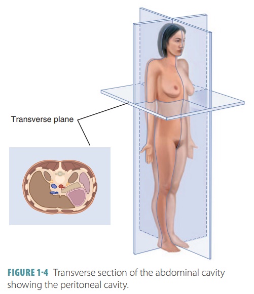
In the abdominopelvic cavity, which extends from the diaphragm to the
pelvis, there are subdi-visions known as the superior abdominal cavity, and the inferior pelvic cavity. The abdominopelvic cavity contains the peritoneal cavity (FIGURE 1-4). This
is a potential space that is lined by a serous membrane called the peritoneum. The peritoneal membranes include
the parietal peritoneum lining the
walls and the visceral peritoneum
covering each organ. Move-ment of the digestive organs, while able to cause
rumbling or gurgling sounds, does not cause damage because the peritoneum
allows them to slide across each other.
The abdominal cavity extends from
the infe-rior surface of the diaphragm to the level of the superior edges of
the pelvis. It contains the liver, stomach, spleen, small intestine, and the
major-ity of the large intestine. These organs are completely or partially
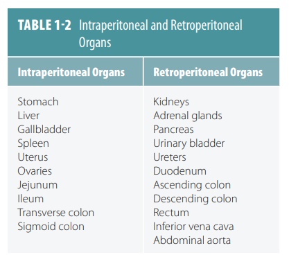
enclosed by the peritoneal cavity. However, certain organs such as the kidney
and pancreas lie in between the peritoneal lining and the muscular abdominal
cavity wall. Organs lying behind the peritoneum are called retroperitoneal. Organs
lying inside the peritoneum are called intraperitoneal. TABLE 1-2 lists intraperitoneal and retroperitoneal organs.
1. List the cavities of the head.
2. Which body cavity will be opened if an incision is made
just inferior to the diaphragm?
3. Identify the subdivisions of the ventral body cavity.
Diagnostic Imaging
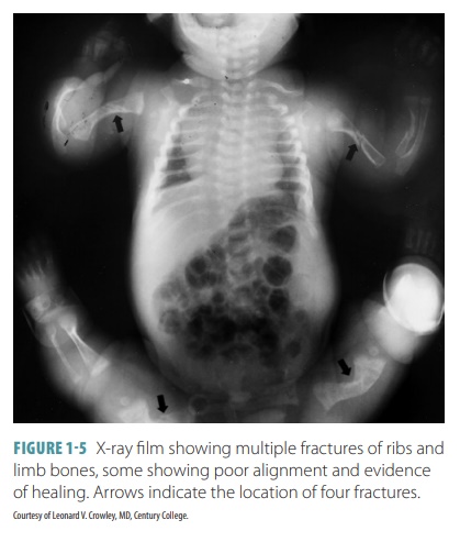
Diagnostic or “medical” imaging
was developed in order to view the internal organs and body structures, in both
normal and abnormal conditions. It began in the first decade of the 1900s when
physicist Wilhelm Roentgen discovered X-rays. Until the 1950s, X-rays were the
exclusive method of imaging available. In its early days, X-rays took much
longer to produce and exposed the patient to significantly higher amounts of
radiation. An example of an X-ray is shown in FIGURE
1-5.
Additional medical imaging
developments are as follows:
■■ The development of fluorescent
screens that were used with special glasses allowed real time viewing of X-ray
images but also exposed physicians to radiation.
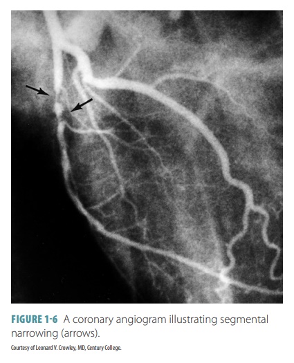
■■Contrast agents barium and
iodine help to improve viewing of the esophagus, stomach, coronary arteries,
and other structures. Examples of procedures that use contrast agents include
intravenous pyelogram and angiogram (FIGURE 1-6).
■■ In 1955, X-ray image
intensifiers allowed movingX-rays to be viewed by using television camerasand
monitors.
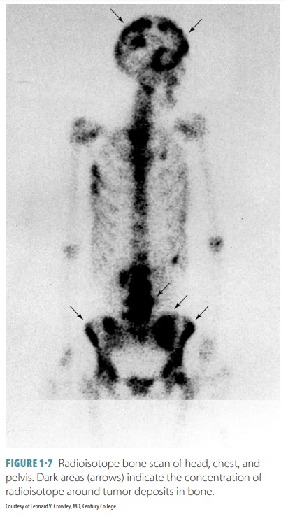
■■ Radionuclide scanning, or
nuclear medicine, wasdeveloped in the 1950s. This type of scan usesspecial
gamma cameras and low level radioactivechemicals introduced into the body,
allowing theevaluation of functional activity of organs. Resultsof nuclear
medicine are recorded as a nuclearisotope scan (FIGURE 1-7).
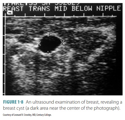
■■ Ultrasound scanning appeared
in the 1960s, using high frequency sound waves to penetrate thebody, bounce off
the internal structures, and thenbe reconstructed into live pictures by a
computer(FIGURE 1-8). Ultrasound is most useful for softtissues and body fluids
and is commonly used toview the gallbladder, urinary bladder, and uterus.
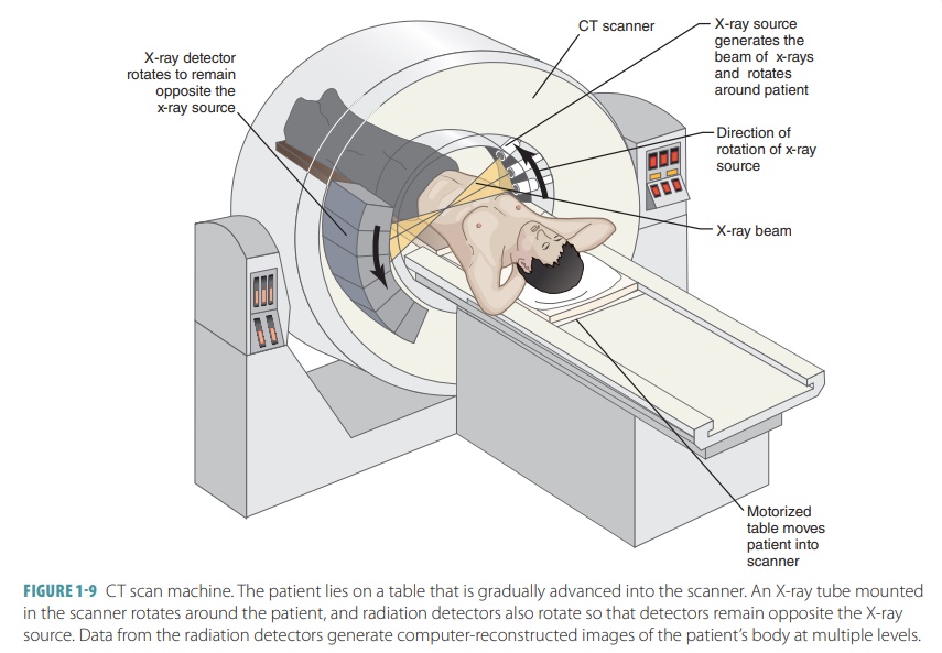
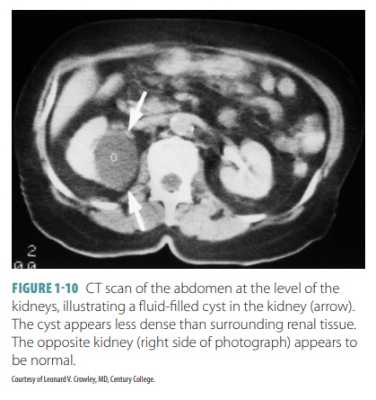
■■ Digital imaging came along in
the 1970s with the development of CT. All preexisting technologies were
upgraded to digital forms. Digital X-ray detectors are replacing previous
analog technologies,allowing better imaging and less healthrisks. CT acquires
an image in less than a secondand instantly reconstructs it. It offers detailed
cross-sectional images of body structures.FIGURE 1-9 shows a CT machine and
FIGURE 1-10shows a CT scan of the abdomen.
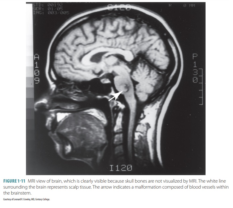
■■ MRI, first offered in 1984,
allows detailed imaging without exposure to radiation (FIGURE 1-11).Images are
produced by displacing protons inatomic nuclei with radiofrequency signals.
However, it cannot be used on a patient who has anymetal implants because of
its extremely powerful magnetization. Also, the person must remain completely
still for a long period of time in asmall, confined space. MRI is often used
for bone,joint, brain, and nerve imaging.
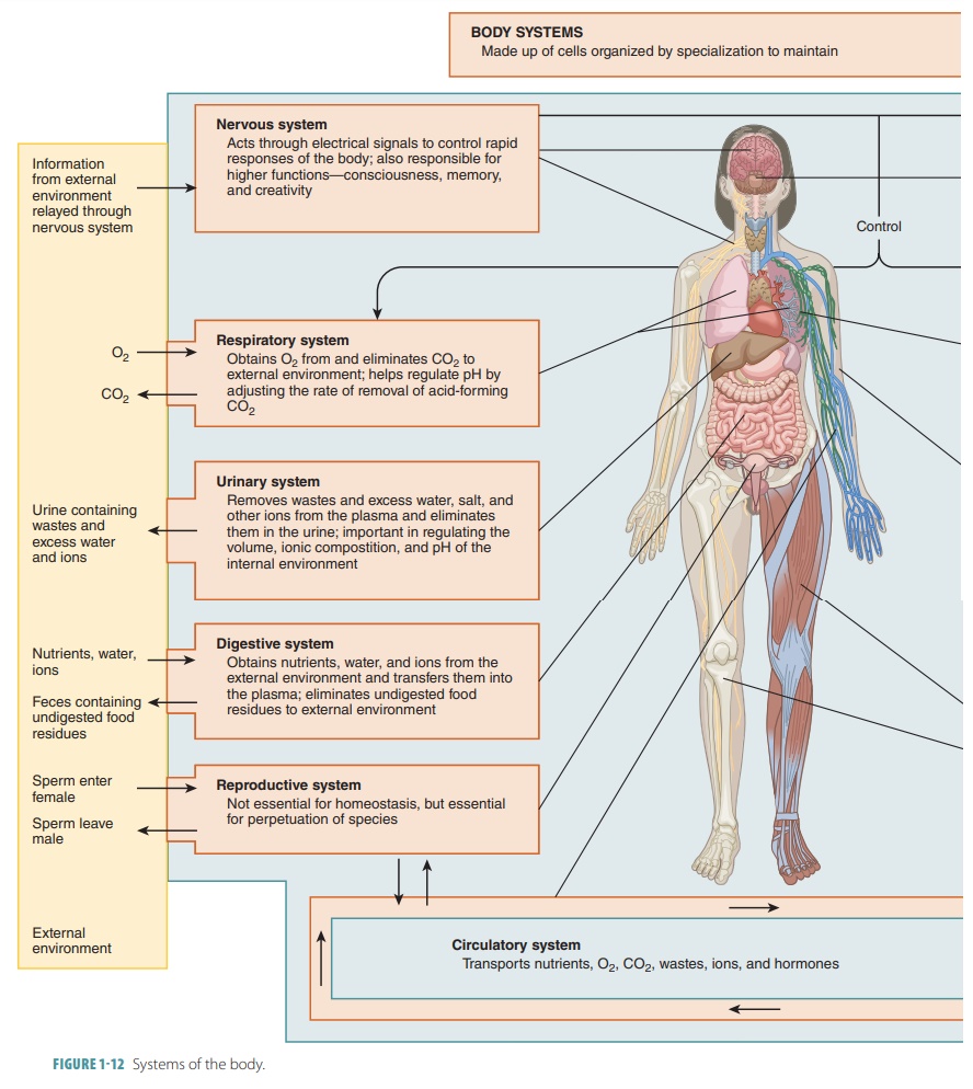
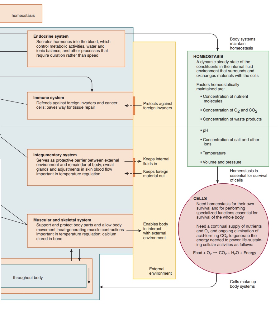
Organ Systems
In each organ system of the human
body, the organs work together to maintain homeostasis. These organ systems
include the integumentary, skeletal, muscular, nervous, endocrine,
cardiovascular, lymphatic, digestive, respiratory, urinary, and reproductive
systems (FIGURE 1-12).
Integumentary System
The integumentary system includes the skin, hair, nails, sebaceous (oil) glands, and sweat glands. The system protects the underlying tissues of the body, assists in the regulation of body temperature, contains various sensory receptors, and manufactures certain substances (such as vitamin D).
Skeletal System
The skeletal system supports and
protects the soft tis-sues of the body and helps the body to move. It con-sists
of bones, which are bound together by ligaments and cartilages. The skeletal
system shields soft tissues and attaches to muscles. The bones also help in
blood formation and provide storage of mineral salts.
Muscular System
The muscular system works with
the skeletal system in helping the body to move. Body parts are moved by muscle
contraction. Posture and body heat are main-tained by the muscular system. The
muscular system also includes the tendons.
Nervous System
The nervous system, along with
the endocrine system, controls and coordinates various organ functions, helping
to maintain homeostasis. The nervous system consists of the brain, spinal cord,
nerves, and sensory organs. Nerve
impulses are electrochemical signals used by nerve cells to communicate
with each other and with the glands and muscles of the body. Certain nerve
cells (called sensory receptors)
detect internal and external changes that affect the body. Other nerve cells
interpret and respond to these stimuli. Additional nerve cells carry impulses
from the brain or spinal cord to the glands and muscles. These nerves are able
to stimulate the muscles to contract and cause the glands to secrete their
products. The characteris-tics of the nervous system include short-term
effects, rapid responses, and very specific responses, as well as a variety of
other responses.
Endocrine System
The endocrine system consists of
hormone-secreting glands. Hormones affect specific target cells, altering their
metabolism. Hormones have a relatively long duration of action compared with
nerve impulses and can last for several days or longer. The endocrine system
also produces a slower response regarding body changes than the nervous system.
The organs of the endocrine system include the hypothalamus (in the brain),
pituitary gland, pineal gland, thyroid gland, parathyroid glands, adrenal
glands, pancreas, and thymus. Other organs with endocrine function include the
ovaries and testes, which are also the parts of the reproductive sys-tem. The
endocrine system can produce effects involv-ing several organs or tissues at
the same time.
Cardiovascular System
The cardiovascular system
includes the heart, blood, arteries, veins, and capillaries. The heart muscle
pumps blood through the arteries, transporting gases, hormones, nutrients, and
wastes. Blood returns to the heart via the veins. Oxygen is carried from the
lungs to the body, and nutrients are carried from the diges-tive system. The
blood also transports biochemicals required for metabolism. Wastes are carried
in the blood from body cells to the excretory organs.
Lymphatic System
The lymphatic system is composed
of the lymphatic vessels, lymph nodes, thymus, spleen, and lymph fluid. It
works with the cardiovascular system, transporting tissue fluid back into the
bloodstream. It also carries specific fats from digestive organs into the
bloodstream. Lymphatic cells (lymphocytes) defend the body against infection.
The lymphatic vessels have two ducts in the chest, known as the thoracic duct
and the right lymphatic duct.
Digestive System
The digestive system takes in
food from outside the body, breaks it down and absorbs the nutrients. It then
excretes wastes from its various processes. The digestive system also produces
certain hormones and works in conjunction with the endocrine system. The
structures of the digestive system include the mouth, teeth, salivary glands,
tongue, esophagus, stomach, liver, gallbladder, pancreas, small intestine,
large intestine, rectum, and anus. The pharynx is part of both the digestive
and respiratory systems.
Respiratory System
The respiratory system takes in
and expels air, exchanging oxygen and carbon dioxide via the lungs and
bloodstream. The structures of the respiratory system include the nose, nasal
cavity, larynx, trachea, bronchi, and lungs. Again, the pharynx is part of both
the respiratory and digestive systems.
Urinary System
The urinary system functions to
remove liquid wastes from the body. It consists of the kidneys, ureters,
urinary bladder, and urethra; it is through the urethra that urine is expelled.
The female urethra is located just above the vagina, while the male urethra
runs through the penis. The kidneys filter wastes from the blood and maintain
electrolyte concentrations. The urinary bladder stores the urine and the
urethra car-ries it to outside the body.
Reproductive System
The reproductive system in
females consists of the ova-ries, uterine tubes, uterus, vagina, clitoris, and
vulva. The female sex cells are called oocytes
or eggs. They are fertilized by male
sex cells (sperm or spermatozoa). When a female is
impregnated, the embryo develops within the uterus. The male reproductive
system includes the scrotum, testes, epididymides, ductusdeferentia, seminal vesicles,
prostate gland, bulbourethral glands, penis, and urethra. Reproduction is the
process of producing offspring. As embryonic cells divide, they grow and
produce new cells, and the pro-cess is continued.
1. Describe
the general functions of the digestive system.
2. List the organs of the respiratory system.
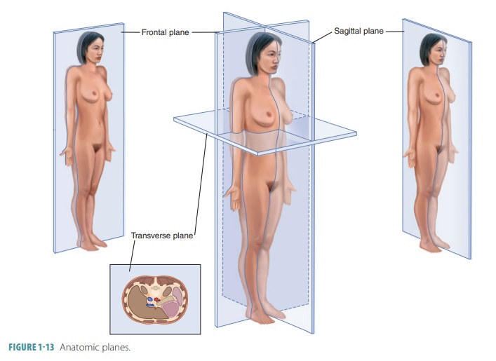
Anatomic Planes
The body can be visually divided into specific areas, called planes. These planes “divide” the body at par-ticular angles and in particular directions (FIGURE 1 -13 and TABLE 1-3). They are also referred to as slices or Sections . There are three sectional planes, as follows:
■■ Transverse (horizontal) plane:
It lies at right anglesto the long axis of the body and divides the bodyinto
superior and inferior portions. A cut in thisplane is called a transverse
(cross) section.
■■ Frontal (coronal) plane: It is
parallel to the longaxis of the body and extends vertically, dividing the body
into anterior and posterior portions.
■■ Sagittal plane: Also parallel
to the long axis ofthe body, this plane, however, divides the body into left
and right portions. A cut passing alongthe midline, dividing the body into
equal leftand right halves, is called a midsagittal (median)section and a cut
parallel to the midsagittal line iscalled a parasagittal section.
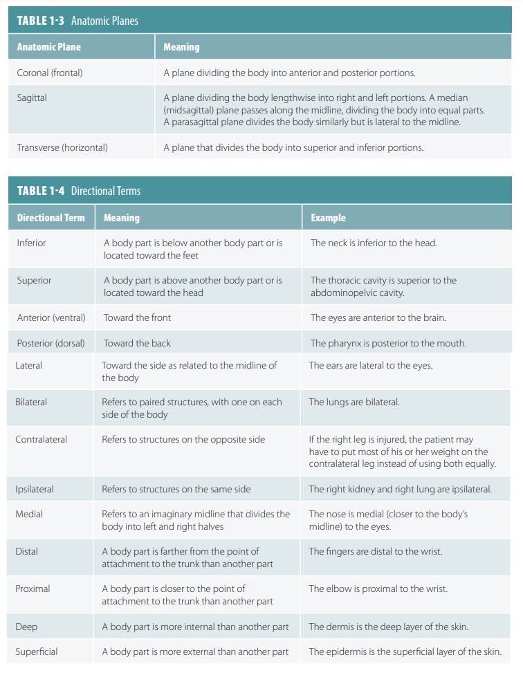
Directional Terms
Directional terms used in the study of anatomy include words that describe relative positions of body parts as well as imaginary anatomical divisions. The term anatomical position describes the body standing erect, facing forward, with the arms held to the sides of the body, palms of the hands facing forward. When the terms right and left are used, they refer to those specific sides of the body when it is in the anatomical position. Important directional terms used in anat-omy are listed in TABLE 1-4.
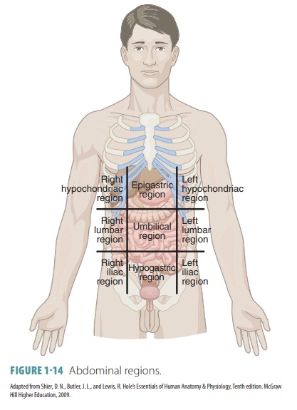
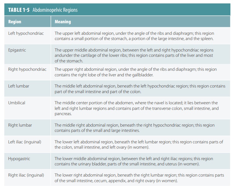
Abdominal Regions
Anatomists have divided the abdomen and pelvisinto nine imaginary
regions that are helpful in identifying the location of particular abdominal
organs.
They are also useful for
describing the location of abdominal pain. FIGURE
1-14 shows the nine abdominal
regions, identified from the left to the right and moving from top to bottom
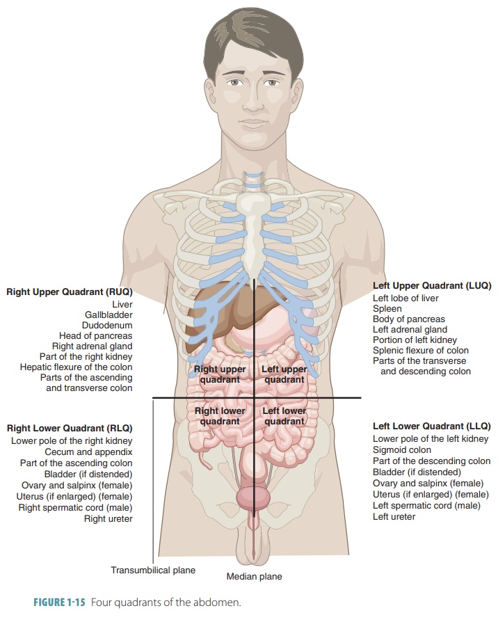
one row at a time. The abdomen may also be divided into four quadrants, as
shown in FIGURE 1-15. TABLE 1-5 explains each abdom-inal region in greater detail.
1. Name
the nine abdominopelvic regions.
2. Describe
the anatomic planes of the body.
3. Describe the term “anatomical position.
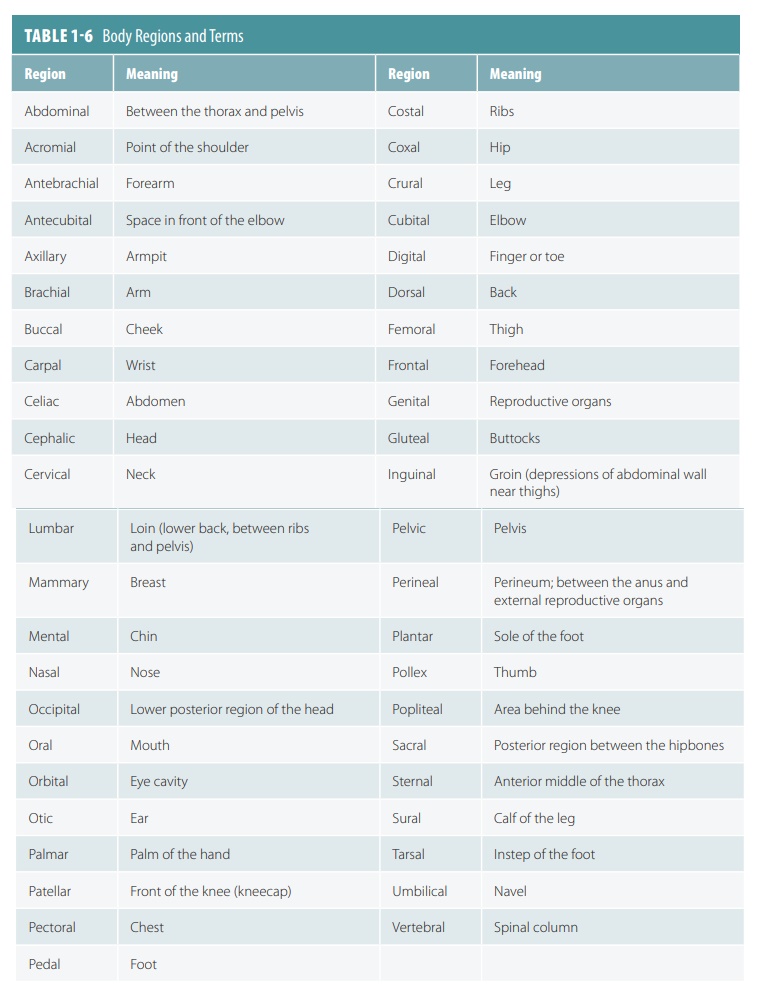
Body Regions
The remainder of the body is
classified into various regions that describe them clinically. For example, carpal tunnel syndrome refers to the
carpal area the wrist where acute pain can occur from the development of this
syndrome. The most common body regions are listed in TABLE 1-6.
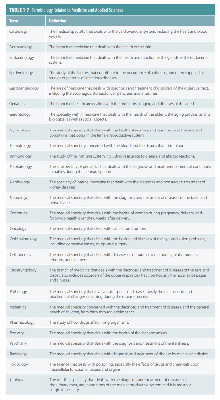
TABLE 1-7 lists terms related to medicine andapplied sciences.