Structure of the Fungal Cell
| Home | | Pharmaceutical Microbiology | | Pharmaceutical Microbiology |Chapter: Pharmaceutical Microbiology : Fungi
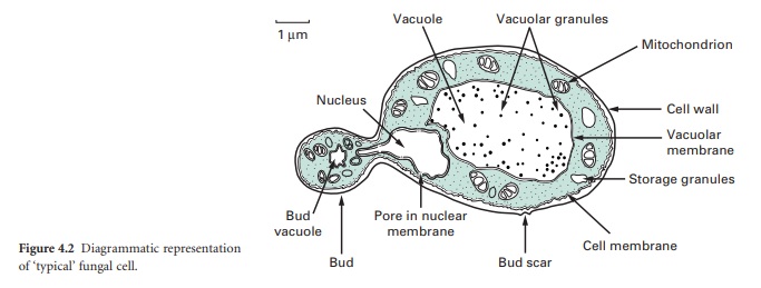
The typical yeast cell is oval in shape and is surrounded by a rigid cell wall which contains a number of structural polysaccharides and may account for up to 25% of the dry weight of the cell wall..
STRUCTURE OF THE FUNGAL CELL
The typical yeast cell
is oval in shape and is surrounded by a rigid cell wall which contains a number
of structural polysaccharides and may account for up to 25% of the dry weight
of the cell wall (see Figure 4.2). Glucan accounts for 50–60%, mannan for 15–23%
and chitin for 1–9% of the dry weight of the wall, respectively, with protein
and lipids also present in smaller amounts. The thickness of the cell wall may
vary during the life of the cell but the average thickness in the yeast C. albicans varies from 100 to 300 nm.
Glucan, the main structural component of the fungal cell wall, is a branched
polymer of glucose which exists in three forms in the cell: β-1,6-glucan, β-1,3-glucan and β1,3,-β-1,6-complexed with chitin. Mannan is a polymer
of the sugar mannose and is found in the outer layers of the cell wall. The
third principal structural component, chitin, is concentrated in bud scars that
are areas of the cell from which a bud has detached. Proteins and lipids are
also present in the cell wall and under some conditions may represent up to 30%
of the cell wall contents. Mannoproteins form a fibrillar layer that radiates
from an internal skeletal layer that is formed by the polysaccharide component
of the cell wall. The innermost layer is rich in glucan and chitin which
provides rigidity to the wall and is important in regulating cell division.
Enzymatic or mechanical
removal of the cell wall leaves an osmotically fragile protoplast which will
burst if not maintained in an osmotically stabilized environment. Incubation of
protoplasts in an osmotically stabilized agar growth medium will allow the re-synthesis
of the wall and the resumption of normal cellular functions. The ability to
generate fungal protoplasts opens the possibility of fusing these under defined
conditions to generate strains with novel biotechnological applications.
The periplasmic space is
a thin region that lies directly below the cell wall. It contains secreted
proteins that do not penetrate the cell wall and is the location for a number
of enzymes required for processing nutrients prior to entry into the cell. The
cell membrane or plasmalemma is located directly below the periplasmic space
and is a phopholipid bilayer which contains phospholipids, lipids, protein and
sterols. The plasmalemma is approximately 10 nm thick and in addition to being
composed of phospholipids also contains globular proteins. The dominant sterol
in fungal cell membranes is ergosterol which is the target of the antifungal
agent amphotericin B. Sterols are important components of the plasmalemma and
represent regions of rigidity in the fluidity provided by the phospholipid
bilayer.
Most of the cell’s
genome is concentrated in the nucleus which is surrounded by a nuclear membrane
which contains pores to allow communication with the rest of the cell (see Figure
4.2 ). The nucleus is a discrete organelle and, in addition to being the
repository of the DNA, also contains proteins in the form of histones. Yeast
chromosomes vary in size from 0.2 to 6 Mb and the number per yeast is also
variable with S. cerevisiae having as
many as 16 while the fission yeast Sch.
pombe has as few as 3. In addition to the genetic material in the nucleus
the yeast cell often has extrachromosomal information in the form of plasmids.
For example, the 2 μm plasmid is present in S. cerevisiae, although its function is
unclear, and there are killer plasmids in the yeast Kluyveromyces lactis which
encode a toxin.
Actively respiring
fungal cells possess a distinct mitochondrion which has been described as the ‘powerhouse’
of the cell (Figure 4.2). The enzymes of the tricarboxylic acid cycle (Krebs’
cycle) are located in the matrix of the mitochondrion while electron transport
and oxidative phosphorylation occur in the mitochondrial inner membrane. The
outer membrane contains enzymes involved in lipid biosynthesis. The
mitochondrion is a semi-independent organelle as it possesses its own DNA and
is capable of producing its own proteins on its own ribosomes which are
referred to as mito-ribosomes.
The fungal cell contains
a vast number of ribosomes which are usually present in the form of polysomes—
lines of ribosomes strung together by a strand of mRNA. Ribosomes are the site
of protein biosynthesis. The system which mediates the export of proteins from
the cell involves a number of membranous compartments including the Golgi
apparatus, the endoplasmic reticulum and the plasmalemma. In addition, the vacuole
is employed as a ‘storage space’ where nutrients, hydrolytic enzymes or
metabolic intermediates are retained until required.
Related Topics
