Veins and Their Branches
| Home | | Anatomy and Physiology | | Anatomy and Physiology Health Education (APHE) |Chapter: Anatomy and Physiology for Health Professionals: Vascular System
1. Explain the superior vena cava and the inferior vena cava. 2. Explain the branches of the brachiocephalic vein. 3. Compare the internal and external jugular veins. 4. Which veins drain the dural sinuses? 1. Explain the branches of the subclavian veins. 2. Define the median cubital vein and its origination. 3. Explain the junctions of the brachial vein. 4. Compare the azygos and hemiazygos veins. 1. Which veins return blood to the right atrium? 2. What is the name of the vein that carries blood from the stomach, intestines, pancreas, and spleen through to the liver? 3. Describe the hepatic portal system. 4. Compare the structures and functions of the superior vena cava with the great saphenous vein. 5. Which vein drains blood from the common iliac veins?
Veins
and Their Branches
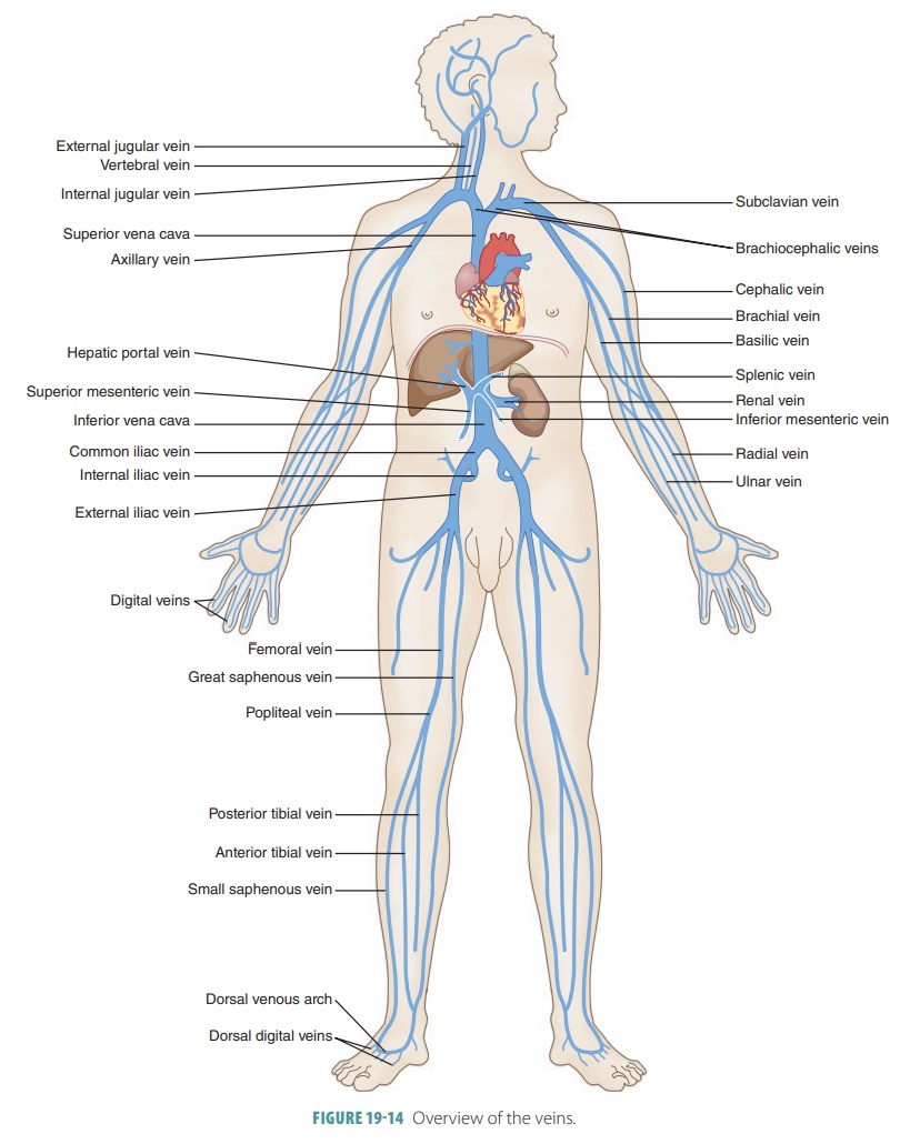
Three major veins return blood from the body to the right atrium: the coronary sinus, superior venacava, and inferior vena cava. The coronary sinusreturns deoxygenated blood from the walls of the heart. The superior vena cava returns blood from the head, neck, thorax (superior to the diaphragm, except the heart wall), and upper limbs. The inferior vena cava returns blood from the abdomen, pelvis, and lower limbs (FIGURE 19-14).
The three major types of veins are the superficial veins,
deep veins, and sinuses. Most superficial veins are larger than the deep veins,
but in the head and trunk the opposite is true. Venous sinuses are mostly found
in the cranial cavity and the heart. The car-diac veins transport blood from
the wall of the heart, returning it via the coronary sinus to the right atrium.
Large
veins have all three tunica layers. Theyinclude the superior and
inferior vena cava and their tributaries inside the thoracic and abdominopelvic
cavities. A thick tunica externa, made of elastic and collagen fibers,
surrounds a thin tunica media. The medium-sized
veins are similar in size to the musculararteries, ranging in internal
diameter between 2 and 9 mm. Their thickest layer is the tunica externa, with
the tunica media being thin and having few smooth muscle cells in relation to
other veins.
Head and Neck Veins
From the head and neck, three pairs of veins collect most
of the draining blood: the external jugu-lar veins, internal jugular veins, and vertebral
veins (FIGURE
19- 15). The more superficial external jugular veins drain blood
mostly from the posterior head and neck, emptying into the subclavian veins.
The larger, deeper internal jugular veins drain blood from the cranial cavity
and the anterior head, neck, and face. The vertebral veins empty into the
brachiocephalic vein. Most extracranial veins have the same names as their
related extracranial arteries. However, their loca-tions and connections are
different.
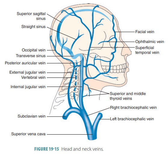
The dural venous sinuses are interconnected, enlarged chambers between the layers of the dura mater. It is here that most veins of the brain drain blood. In the falx cerebri, which is located down between the cere-bral hemispheres, are found the superior and inferiorsagittal sinuses. Mostinferior cerebral veinsconvergeto form the great cerebral vein. Posteriorly, the inferior sagittal sinus drains into the straight sinus. Then, the superior sagittal and straight sinuses drain into the transverse sinuses. These sinuses are located insideshallow grooves on the occipital bone’s internal sur-face. They drain into the sigmoid sinuses, which are S-shaped. The sinuses then become the internal jugularveins when they leave the skull via the jugular foramen.The cavernous sinuses receive blood from the ophthalmic veins of the orbits and facial veins. These sinuses flank the sphenoid body and drain the nose and upper lip region. On the way to the face and orbits, the internal carotid artery; cranial nerves III, IV, and VI; and part of cranial nerve V run through the cavernous sinus.
External Jugular Veins
Superficial scalp and face structures supplied by the
external carotid arteries are drained by the left and right external jugular veins. They anastomose often, and an
amount of superficial drainage from them also enters into the internal jugular
veins. The external jug-ular veins descend through the lateral neck, passing
obliquely over the sternocleidomastoid muscles. They finally empty into the
subclavian veins. The superficial head and neck veins converge to form the temporal,facial, and maxillary veins. The temporal and maxil-lary veins drain into the external jugular vein.
Vertebral Veins
The vertebral veins
are not similar to the vertebral arteries in that they do not serve most areas
of the brain. They drain the spinal cord, cervical vertebra, and some of the
neck’s small muscles. They are located inferiorly through the cervical
vertebra’s transverse foramina. The vertebral veins join the brachiocephalic
veins at the root of the neck.
Internal Jugular Veins
Most of the blood draining from the brain is received by the
two internal jugular veins, the
largest paired veins that drain the head and neck, emerging from the dural
venous sinuses. The internal jugular veins exit the skull through the jugular foramina and descend through the
neck beside the internal carotid arteries. Further downward, they receive blood
from certain deep face and neck veins that are branches of the facialand superficial temporal veins. On either side of thebase of the
neck, each internal jugular vein joins its subclavian vein, forming a brachiocephalic vein. Then, as previously described, the
two brachioce-phalic veins join and form the superior vena cava.
1. Explain the superior vena cava and the inferior vena cava.
2. Explain the branches of the brachiocephalic vein.
3. Compare the internal and external jugular veins.
4. Which
veins drain the dural sinuses?
Upper Limb and Thoracic Veins
As previously described, the deep upper limb veins follow
their related arteries’ paths and therefore have the same names. Most are
paired veins flanking their related artery, except for the largest veins.
Because the superficial upper limb veins are larger than the deep veins, they
can be easily seen beneath the skin.
Deep Upper Limb Veins
The radial and ulnar veins are the most distal deep veins of
the upper limbs. From the hand, the deep
and superficial venous palmar arches empty into the radial and ulnar veins of the forearm. Then theyjoin, forming the brachial vein of the arm. At the axilla,
the brachial vein becomes the axillary
vein. Then, at the level of the first rib, it becomes the subclavian vein.
Superficial Upper Limb Veins
The dorsal venous
network begins the superficial veins of the upper limbs. It is a plexus of
superficial veins located in the dorsum of the hand. The digi-tal veins empty into the
superficial and deep palmar veins of
the hand, which join to form the palmar
venous arches. The dorsal venous network drainsinto two major superficial
veins in the distal fore-arm, the cephalic
and basilic veins, which have many anastomoses as they continue upward. As
it con-tinues in this manner, the cephalic
vein encircles the radius and continues to the shoulder by
moving up the lateral superficial aspect of the arm. At the shoulder, the
cephalic vein moves into the groove between the deltoid and pectoralis
muscles. Here, it joins the axillary vein. FIGURE 19-16 shows the upper limb and thoracic veins.
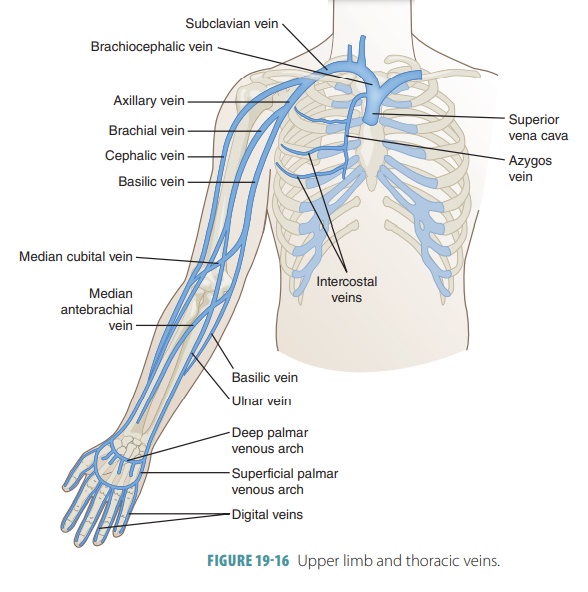
The basilic
vein continues along the forearm’s posteromedial aspect,
crossing the elbow, and join-ing the brachial vein in the axilla to form the
axillary vein. The medial cubital vein
connects the basilic and cephalic veins at the anterior aspect of the elbow.
The median antebrachial vein,
between the radial andulnar veins, usually ends at the elbow as it enters
either the basilic or cephalic vein. The radial
and ulnar veins arise from the deep
palmar veins of the hand.
Azygos Vein
The azygos
vein is located against the right side of the vertebral column.
It begins in the abdomen, via the right
ascending lumbar vein. This vein drains mostof the right abdominal
cavity wall. The azygos vein also begins from the right posterior intercostal veins,
except for the first of these. These veins drain the mus-cles of the chest. At
the T4 level, the azygos vein arches over the great vessels that supply the
right lung. The azygos vein empties into the superior vena cava. Other veins
that drain the abdomen include the lumbar, gonadal, hepatic, renal, adrenal,
and phrenic veins.
Hemiazygos Vein
The hemiazygos vein
ascends on the vertebral column’s left side, emerging from the left ascending lumbar vein and the 9th
to 11th posterior intercostal veins.
The hemia-zygos vein is similar to the inferior portion of the azygos and superficial venous palmar arches empty
into the radial and ulnar veins of
the forearm. Then theyjoin, forming the brachial
vein of the arm. At the axilla, the brachial vein becomes the axillary vein. Then, at the level of the first
rib, it becomes the subclavian
vein.
Superficial Upper Limb Veins
The dorsal venous
network begins the superficial veins of the upper limbs. It is a plexus of
superficial veins located in the dorsum of the hand. The digi-tal veins empty into the
superficial and deep palmar veins of
the hand, which join to form the palmar
venous arches. The dorsal venous network drainsinto two major superficial
veins in the distal fore-arm, the cephalic
and basilic veins, which have many anastomoses as they continue upward. As
it con-tinues in this manner, the cephalic
vein encircles the radius and continues to the shoulder by
moving up the lateral superficial aspect of the arm. At the shoulder, the
cephalic vein moves into the groove between the deltoid and pectoralis
muscles. Here, it joins the axillary vein. FIGURE 19-16 shows the upper limb and thoracic veins.
The basilic vein continues along the forearm’s posteromedial aspect, crossing the elbow, and join-ing the brachial vein in the axilla to form the axillary vein. The medial cubital vein connects the basilic and cephalic veins at the anterior aspect of the elbow. The median antebrachial vein, between the radial andulnar veins, usually ends at the elbow as it enters either the basilic or cephalic vein. The radial and ulnar veins arise from the deep palmar veins of the hand.
Azygos Vein
The azygos
vein is located against the right side of the vertebral column.
It begins in the abdomen, via the right
ascending lumbar vein. This vein drains mostof the right abdominal
cavity wall. The azygos vein also begins from the right posterior intercostal veins,
except for the first of these. These veins drain the mus-cles of the chest. At
the T4 level, the azygos vein arches over the great vessels that supply the
right lung. The azygos vein empties into the superior vena cava. Other veins
that drain the abdomen include the lumbar, gonadal, hepatic, renal, adrenal,
and phrenic veins.
Accessory Hemiazygos Vein
The hemiazygos vein
ascends on the vertebral column’s left side, emerging from the left ascending lumbar vein and the 9th
to 11th posterior intercostal veins.
The hemia-zygos vein is similar to the inferior portion of the azygos
1. Explain the branches of the subclavian veins.
2. Define the median cubital vein and its origination.
3. Explain the junctions of the brachial vein.
4. Compare
the azygos and hemiazygos veins.
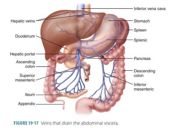
Hepatic Portal System
The pathway of blood flow from the gastrointestinal tract
and spleen to the liver via the portal vein and its tributaries is called the hepatic portal circulation. The hepatic portal system is made up of a series of vessels
and contains two distinct capillary beds lying between the arterial supply and
the final venous drainage. In this system the initial capillary beds are found
in the stomach and intestines. They drain into vessels of the hepatic portal
vein, and the blood is then carried to a second capillary bed inside the liver.
The hepatic portal vein is short,
starting at the L2 level. A group of vessels from the stomach and pancreas
contributes to the hepatic portal system. However, there are three major
vessels: the superior mesenteric vein, splenic vein, and inferior mesenteric
vein.
The superior
mesenteric vein drains all the small intestine, the ascending and
transverse regions of the large intestine, and the stomach. The splenicvein collects blood from
parts of the stomach andpancreas as well as all of the spleen. It joins the
supe-rior mesenteric vein, forming the hepatic portal vein. The inferior mesenteric vein drains the
distal large intestine and rectum. It joins the splenic vein just prior to
its uniting with the superior mesenteric vein (FIGURE 19-17 ) . The hepatic portal vein receives blood from the left
and right gastric veins of the
stomach and from the cystic vein of
the gallbladder.
Pelvic and Lower Limb Veins
Most of the pelvic and lower limb veins also have the same
names as their accompanying arteries. Veins that drain blood from the lower
limbs are also subdivided, similar to those of the upper limbs, into deep and
super-ficial groups (FIGURE
19-18). Many of these veins are double. The two superficial saphenous veins are where
varicosities often occur, because they are not supported very well by
surrounding tissues. In coronary bypass operations the great saphenous vein is often excised and used as a
replacement blood vessel. The gonadalveins
drain the ovaries in females and testicles in males.
Deep Veins
The posterior tibial vein ascends deep in the calf muscle after it forms via the union of the medial andlateral plantar veins. The posterior tibial vein receivesblood from the fibular (peroneal) vein. The superior continuation of the dorsalis pedis vein of the foot is the anterior tibial vein. It unites, at the knee, with the posterior tibial vein, forming the popliteal vein across the back of the knee. The popliteal vein emerges from the knee to become the femoral vein, draining the deep thigh structures. The femoral vein receives blood from the great saphenous, deep femoral, and femoral circumflex veins. As it enters the pelvis, the femoral vein becomes the external iliac vein. In the pelvis this vein joins the internal iliac vein, becom-ing the common iliac vein. The internal iliac veins are arranged very similar to the arrangement of the inter-nal iliac arteries.
Superficial Veins
From the dorsal venous
arch of the foot, the great andsmall
saphenous veins emerge, anastomosing oftenwith each other as well as the
nearby deep veins. The longest vein in the body is the great saphenous vein. It runs from the foot to the femoral vein,
distal to the inguinal ligament. It travels superiorly along the medial aspect
of the leg to the thigh. The small
saphe-nous vein runs from the foot to the knee. It is locatedalong the
lateral aspect of the foot, moving through the deep fascia of the calf muscles.
The small saphe-nous vein drains the calf muscles and empties into the
popliteal vein. The plantar vein collects blood from the capillaries in the
sole of the foot.
1. Which veins return blood to the right atrium?
2. What is the name of the vein that carries blood from the
stomach, intestines, pancreas, and spleen through to the liver?
3. Describe the hepatic portal system.
4. Compare the structures and functions of the superior vena cava with the great saphenous vein.
5. Which vein drains blood from the common iliac veins?
Related Topics