Tissues of the Human Body
| Home |Chapter: HAP - Tissues of the Human Body
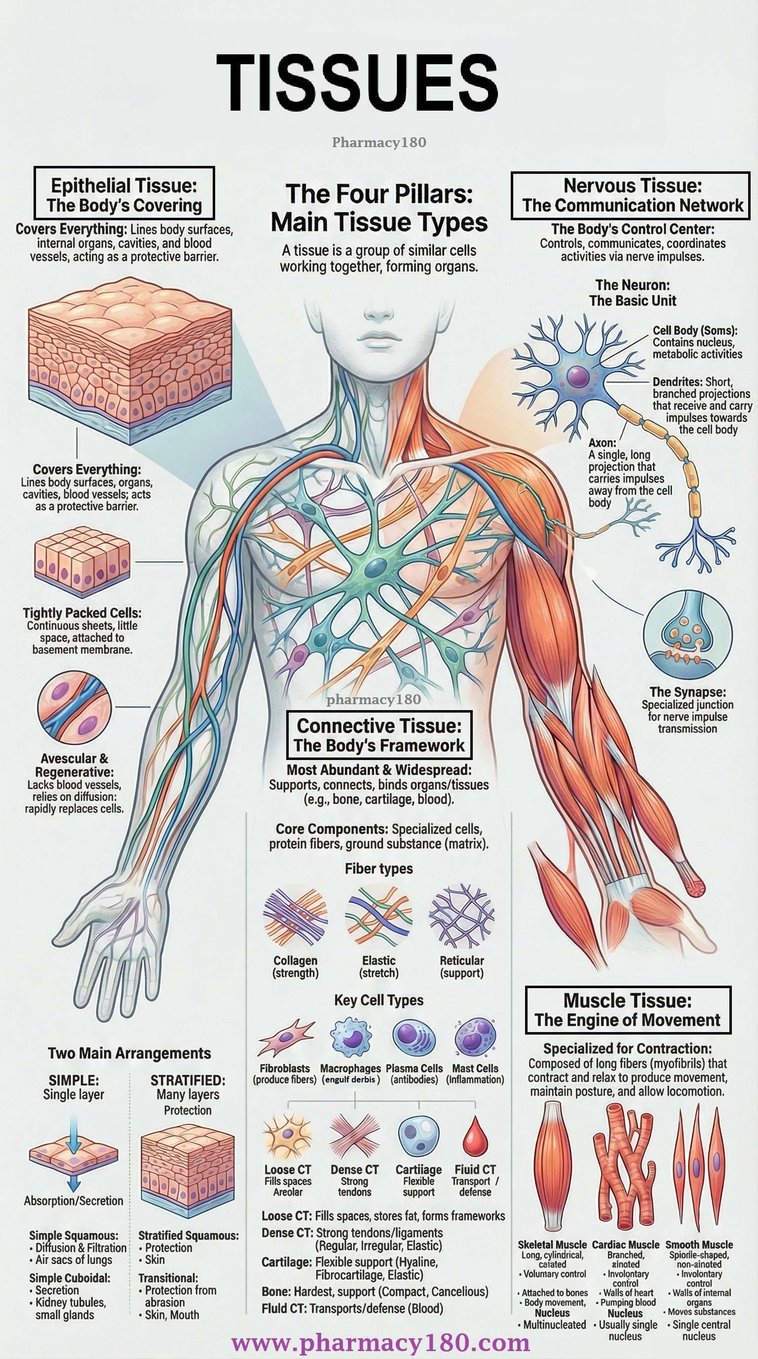
A tissue is a group of cells that have similar structure and work together to perform a specific function.
TISSUES
OF THE HUMAN BODY
TISSUE
A
tissue is a group of cells that have similar structure and work together
to perform a specific function.
Different tissues may be:
- Hard (e.g., bone)
- Semisolid (e.g., fat)
- Liquid (e.g., blood)
Tissues combine to form organs such
as the stomach, heart, lungs, and brain.
The study of tissues is called Histology.
Types of Tissue
The human body contains four main types
of tissues, each performing unique roles:
|
TISSUE TYPE |
MAIN FUNCTION |
|
Epithelial Tissue |
Protection, secretion, absorption |
|
Connective Tissue |
Support, binding, packing |
|
Muscle Tissue |
Movement and locomotion |
|
Nervous Tissue |
Control and coordination |
These four tissues form the foundation of
all organs in the body.
I. EPITHELIAL
TISSUE
Epithelial tissue covers:
- Body
surfaces
- Internal
organs
- Body
cavities
- Blood
vessels
- Glandular
structures
Thus, epithelial tissue acts as a protective
covering and also forms the functional units of glands.
Characteristics of Epithelial Tissue
- Cells
form continuous sheets
– Tightly packed like tiles, with very little space between them. - Apical
Surface
– The top (free) surface faces a cavity or the outside of the body. - Basement
Membrane
– The lower surface attaches to underlying connective tissue. - Avascular
– Contains no blood vessels; nutrients diffuse from nearby connective tissue. - Rapid
regeneration
– Repairs and replaces cells quickly due to constant wear and tear.
Functions of Epithelial Tissue
- Protects
the body from friction, dehydration, and injury
- Allows
selective exchange of chemicals
- Secretes
hormones into the bloodstream
- Produces
sweat, mucus, enzymes, and other secretions
TYPES OF EPITHELIAL TISSUE
Epithelial tissues are classified based on:
1. Based on Arrangement of Layers
i) Simple epithelium
ii) Stratified epithelium
i)
Simple Epithelium – Types
- Simple
squamous epithelium
- Simple
cuboidal epithelium
- Simple
columnar epithelium
- Simple
ciliated epithelium
- Glandular
epithelium
Glandular Epithelium – Types
·
Unicellular
glands
·
Multicellular
glands
o Exocrine glands
o Endocrine glands
ii)
Stratified Epithelium – Types
- Stratified
squamous epithelium
- Keratinised
stratified squamous epithelium
- Non-keratinised
stratified squamous epithelium
- Stratified
cuboidal epithelium
- Stratified
columnar epithelium
- Transitional
epithelium
2. Based on Shape of Cells
- Squamous
epithelium (flat)
- Cuboidal
epithelium (cube-like)
- Columnar
epithelium (tall)
- Ciliated
epithelium
- Glandular
epithelium
SIMPLE EPITHELIUM
Cells
are arranged in a single layer.
Their thinness makes them ideal for absorption, filtration, secretion, and
diffusion.
Types of Simple Epithelium
1. Simple Squamous Epithelium
- Single
layer of flat, thin cells
- Allows
easy diffusion of gases and nutrients
Location:
- Air sacs
of lungs
- Lining of
blood & lymph vessels
- Heart
lining
Function:
- Lubrication
- Diffusion
- Filtration
2. Simple Cuboidal Epithelium
- Single
layer of cube-shaped cells
- Often
involved in secretion and absorption
Location:
- Kidney
tubules
- Small
gland ducts
Function:
- Secretion
- Absorption
3. Simple Columnar Epithelium
- Tall,
pillar-shaped cells
- May
contain microvilli or goblet cells (mucus-secreting)
Location:
- Digestive
tract lining (non-ciliated)
- Uterine
tubes, uterus, bronchi (ciliated)
Function:
- Absorption
- Secretion
of mucus and enzymes
4. Simple Ciliated Epithelium
- Columnar
cells with cilia on the free surface
- Cilia
move substances like mucus or ovum
Location:
- Trachea,
upper respiratory tract
- Fallopian
tubes
- Spinal
cord canal
Function:
- Moves
mucus or reproductive cells
5. Glandular Epithelium
- Specialized
for secretion
- Made of
cuboidal or columnar cells containing secretory granules
Types of Glands:
|
TYPE |
DESCRIPTION |
|
Unicellular |
Goblet cells producing mucus |
|
Multicellular |
Exocrine (with ducts), Endocrine
(ductless) |
Exocrine glands: Secrete enzymes, sweat, saliva
Endocrine glands: Secrete hormones directly into blood
STRATIFIED EPITHELIUM
Also
called compound epithelium.
It has many layers of cells and mainly provides protection.
It usually lacks a distinct basement
membrane due to multiple layers.
Types of Stratified Epithelium
1. Stratified Squamous Epithelium
Has many layers:
- Bottom
layers → cuboidal/columnar
- Top
layers → flattened (squamous)
Two forms:
a) Keratinised Stratified Squamous
Epithelium
- Contains keratin,
a tough waterproof protein
- Found on dry
surfaces exposed to friction
Location: Skin, hair, nails
b) Non-keratinised Stratified Squamous
Epithelium
- Moist
surfaces
- Protects
from friction and prevents drying out
Location:
- Mouth
- Pharynx
- Esophagus
- Vagina
- Conjunctiva
Function (both forms):
- Protection
from wear and tear
2. Stratified Cuboidal Epithelium
- Two or
more layers
- Apical
layer contains cuboidal cells
Location:
- Sweat
gland ducts
- Male
urethra
- Uterus
and anus
Function:
- Protection
- Secretion
- Absorption
3. Stratified Columnar Epithelium
- Several
layers
- Apical
layer contains columnar cells
Location:
- Large
excretory gland ducts
- Conjunctiva
of the eye
- Parts of
urethra
Function:
- Protection
- Secretion
4. Transitional Epithelium
- Cells are
pear-shaped and stretchable
- Looks
cuboidal when relaxed but squamous when stretched
Location:
- Urinary
bladder
- Ureters
Function:
- Allows
stretching
- Protects
underlying tissues
II. CONNECTIVE
TISSUE
Connective tissue is the most abundant
and widely distributed tissue in the human body.
It supports, connects, and binds various organs and tissues together.
Examples include: cartilage, bone,
adipose tissue, areolar tissue, and blood.
Connective tissue contains:
- Cells
- Fibres
- Ground
Substance (Matrix)
These three components give strength,
elasticity, and support to the tissue.
Functions of Connective Tissue
- Provides mechanical
support to organs
- Connects
and binds tissues
- Acts as a
medium for exchange of nutrients and waste
- Stores
energy (fat tissue)
- Provides insulation
- Offers defence
– barriers, antibodies, phagocytosis
Composition of Connective Tissue
Connective tissue has fibres, cells,
and ground substance.
FIBRES
1. Collagen Fibres
- Thick
& strong
- Provide tensile
strength
- Most
abundant fibre in connective tissue
2. Elastic Fibres
- Made of elastin
- Stretch
and recoil easily
- Found in
skin, lungs, arteries
3. Reticular Fibres
- Thin,
branched fibres
- Form
delicate networks
- Provide
support in soft organs (spleen, lymph nodes)
CELLS
1. Fibroblasts
- Most
common cell
- Produce
fibres and ground substance
2. Macrophages
- “Big
eaters”
- Perform phagocytosis
of foreign bodies and microbes
- Two
types:
- Wandering
macrophages – move
to infection sites
- Fixed
macrophages – remain
in organs (lungs, spleen)
3. Plasma Cells
- Derived
from B-lymphocytes
- Produce antibodies
- Found in
respiratory and digestive tract, lymph nodes
4. Mast Cells
- Secrete histamine,
important during inflammation
- Dilate
blood vessels
TYPES OF CONNECTIVE TISSUE
Connective tissue is broadly classified
into:
- Loose
Connective Tissue
- Dense
Connective Tissue
- Cartilage
Tissue
- Bone
Tissue
- Blood
(Fluid Connective Tissue)
1. LOOSE CONNECTIVE TISSUE
Cells and fibres are loosely arranged in a
semi-fluid matrix.
a) Areolar Tissue
- Most
widely distributed
- Contains
all types of fibres and cells
- Fills
spaces between organs
Location:
Under the skin, between muscles, around blood vessels and nerves
Functions:
- Strength
- Flexibility
- Support
b) Adipose Tissue
- Made of adipocytes
(fat cells)
- Stores
fat as a single large droplet
Location:
Under the skin, around heart and kidneys, yellow bone marrow
Functions:
- Prevents
heat loss (insulation)
- Energy
storage
- Protection
of organs
- Gives
shape to body
c) Reticular Tissue
- Contains
reticular fibres and reticular cells
- Forms a
supportive network (stroma)
Location:
Liver, spleen, lymph nodes, red bone marrow
Functions:
- Forms
structural framework
- Filters
worn-out blood cells (spleen)
- Removes
microbes (lymph nodes)
2. DENSE CONNECTIVE TISSUE
Contains closely packed fibres and fewer
cells.
a) Dense Regular Connective Tissue
- Fibres
arranged in a parallel pattern
- Very
strong
Location:
- Tendons
(muscle → bone)
- Ligaments
(bone → bone)
Function:
- Provides
strong attachments
b) Dense Irregular Connective Tissue
- Fibres
arranged in irregular patterns
- Can
withstand tension from multiple directions
Location:
- Dermis of
skin
- Organ
capsules
c) Elastic Connective Tissue
- Contains
freely branching elastic fibres
- Yellowish
in appearance
Location:
- Lungs
- Elastic
arteries
- Vocal
cords
- Bronchial
tubes
Function:
- Allows
stretching and elasticity of organs
3. CARTILAGE TISSUE
Cartilage is firmer than other
connective tissues but more flexible than bone.
Cells called chondrocytes lie in spaces called lacunae.
Types of Cartilage
a) Hyaline Cartilage
- Most
common type
- Glassy,
bluish-white appearance
- Chondrocytes
arranged in small groups
Location:
Nose, trachea, bronchi, rib ends, fetal skeleton
b) Fibrocartilage
- Many
collagen fibres
- Very
strong and shock-absorbing
Location:
Intervertebral discs, pubic symphysis
c) Elastic Cartilage
- Yellow
elastic fibres
- Most
flexible type
Location:
External ear, epiglottis
4. BONE TISSUE
Bone
is the hardest connective tissue.
Its strength comes from:
- Calcium
salts (calcium phosphate & carbonate)
- Collagen
fibres
Components of Bone
Bone consists of:
- Periosteum
- Compact
bone
- Cancellous
bone
- Bone
marrow
Microscopic Structure
|
STRUCTURE |
DESCRIPTION |
|
Lacunae |
Spaces containing osteocytes (mature bone
cells) |
|
Canaliculi |
Tiny canals connecting lacunae |
|
Lamellae |
Concentric layers around central canal |
|
Haversian Canal |
Contains blood vessels and nerves |
Functions of Bone
- Provides
structural support
- Protects
organs
- Stores
calcium
- Produces
blood cells in red bone marrow
5. BLOOD (FLUID CONNECTIVE TISSUE)
Blood is considered a connective tissue
because it:
- Has cells
suspended in a matrix (plasma)
- Transports
materials throughout the body
Components of Blood
- RBCs
(Erythrocytes)
- WBCs
(Leukocytes)
- Platelets
- Plasma
(matrix)
Functions
- Transport
of gases, nutrients, hormones
- Defence
against infection
- Clotting
- Regulation
of body temperature
III. MUSCLE
TISSUE
Muscle
tissue contains long fibres called myofibrils.
They contract and relax in response to stimulation.
Functions
- Produce
movement
- Maintain
posture
- Allow
locomotion
- Provide
protection
- Help
adjust body position
TYPES OF MUSCLE TISSUE
1. Skeletal Muscle Tissue
- Long,
cylindrical, striated fibres
- Multinucleated
- Under voluntary
control
Location: Attached to bones
Function: Movement, posture, facial expressions
2. Cardiac Muscle Tissue
- Striated,
branched cells
- Connected
by intercalated discs
- Involuntary
control
Location: Walls of the heart
Function: Pumps blood throughout the body
3. Smooth Muscle Tissue
- Spindle-shaped
cells
- No
striations
- Single
central nucleus
- Involuntary
Location:
- Walls of
blood vessels
- Digestive
tract
- Uterus
- Bladder
- Respiratory
tract
- Iris of
eye
Functions:
- Digestion
and movement of food
- Urine
flow regulation
- Control
of diameter of blood vessels
- Movement
of reproductive cells
IV. NERVOUS
TISSUE
Nervous tissue is responsible for control
and communication in the body.
The basic structural and functional unit is
the neuron.
A neuron has three main parts:
1. Cell Body (Soma)
- Contains
nucleus
- Controls
metabolic activities
- Gives
rise to axon and dendrites
2. Axon
- Longest
part of neuron
- Carries
impulses away from the cell body
- May be
covered by:
- Myelin
sheath (fatty
insulation)
- Neurilemma (outer covering)
Breaks in the myelin sheath are called Nodes
of Ranvier.
3. Dendrites
- Short,
branched projections
- Carry
impulses towards the cell body
Synapse
A synapse is the junction between
the axon of one neuron and the dendrite of another.
Location
Brain, spinal cord, peripheral nerves
Functions of Nervous Tissue
- Generates
and transmits nerve impulses
- Responds
to internal and external stimuli
- Communicates
information throughout the body
- Coordinates
body activities
- Produces
neurotransmitters
- Provides
insulation and support
