Nutrition, Cultivation and Isolation of Viruses
| Home | | Pharmaceutical Microbiology | | Pharmaceutical Microbiology |Chapter: Pharmaceutical Microbiology : Nutrition, Cultivation and Isolation of microorganisms : Bacteria-Actinomycetes-Fungi-Viruses
The world has broadly witnessed by 1900 and accepted generally that severlal of the recognized dreadful human ailments were duly caused by various microorganisms.
VIRUSES
The world
has broadly witnessed by 1900 and accepted generally that severlal of the
recognized dreadful human ailments were duly caused by various microorganisms.
However, the first and foremost evidence of viruses responsible for causing
human disease came into notice in 1892 when Iwanowski rightly demonstrated that
the cell-free extracts of the diseased
tobacco leaves passed through the bacteria-proof filters may ultimately
cause disease in the ‘healthy plants’.
Furthermore, such cell-free filtrates when cultured upon the bacterial growth media they eventually
exhibited practically little growth
thereby suggesting that the said filtrates
contained the actual disease causing
agents that are other than microorganisms. Martinus Beijernick, another
scientist reconfirmed the excellent epoch making findings of Iwanowski.
Twort and
d’Herrelle (1915) individually showed the ‘glassy
phenomenon’ present very much in the microorganisms when it was observed
clearly and distinctly that the bacterial cells might be adequately infected
with and duly destroyed by the filterable
agents, which in turn caused various serious diseases both affecting the animal kingdom and the plant kingdom. Later on, these disease
producing filterable agents are known as bacteriophages
(i.e., the bacteria-eaters).
Wendell M
Stanley (1935), an American Chemist, first and foremost isolated the tobacco mo-saic virus (genus Tobamovirus) thereby making it
possible to perform the chemical as well
as struc-tural studies on a purified
virus. Interestingly, almost within the same time, the invention of the elec-tron microscope took place which
eventually made it quite possible to visualize the said viruses for the first time.
The
galloping advancement and progress in the in-depth studies on the viruses across the globe based duly
upon the latest molecular biology
techniques in the 1980s and 1990s have remarkably led to the discovery of
the new dreadful human viruses. In the year 1989, the world has duly
acknowledged the discovery of Hepatitis
C virus, and Pestivirus, which
specifically causes acute pediatric
diarrhoea. The year 1993, critically observed the outbreak of a Hantavirus
infection occurring exclusively in the South-western USA, which essentially
possesses the potential for new infections to emerge at any time. Hantavirus disease refers to the acute
ailment related to respirator disease and may even prove fatal.
1. Bacteriophages
Bacteriophages designated the ‘last group of viruses’ which were
duly recognized and best characterized.
As to date one may have the evidence for the presence of such disease producing
agents that are found to be even smaller in size than the viruses, and termed as viriods.
They usually consist of the nucleic
acids (i.e., DNA and RNA)
exclusively.
Example : The spindle tuber disease of potatoes is a glaring example of a
specific disease invariably caused
by the viriods.
A good
number of bacteriophages infecting various microorganisms have now been
duly iso-lated, characterized, and
recognized. The following Table 5.2. records the variuos bacteriophages, host(s), particle dimensions (viz., head and tail in nm),
structure, and composition adequately.
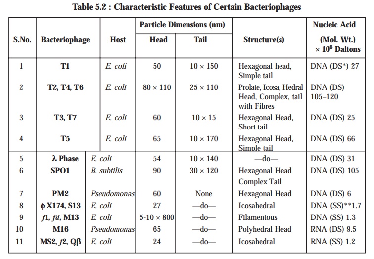
*DS = Double Stranded ; **SS = Single Stranded ;
[Adapted
From : Tauro P et al., An Introduction to Microbiology, New
Age International, New Delhi, 2004].
Viral Species : A viral species may be defined as ‘a group of viruses essentially sharing the same genetic information
and ecological niche.
It is,
however, pertinent to state here that the particular epithets for viruses have not yet been established completely,
thereby logically and emphatically the viral
species are duly designated by such common
descriptive nomenclatures as : human
immunodeficiency virus (HIV), with subspecies duly indicated by a number (HIV-1).
Standardization
of the ‘viral nomenclature’ is now
in an active and progressive stage ; and as such the following specific
criteria are being adopted in the latest textbooks and literature alike, namely
:
·
New viral family
·
Genus names
·
Common species names
·
Common names are expressed in regular type viz., herpes simplex virus
·
Genus names are now usually capitalized and italicized viz.,
Simplexvirus.
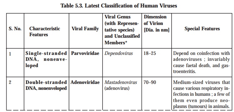
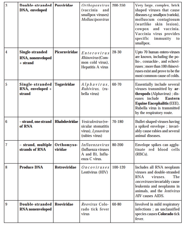
Table
5.3. records a comprehensive summary
of the latest classification of viruses
that invari-ably infect the human beings.
Table 5.3. Latest Classification of Human Viruses
2. Growth of Bacteriophages in the Laboratory
It is
practically possible to grow the bacteriophages
in two different manners, namely :
(a) In suspensions of organisms in liquid media, and
(b) In bacterial cultures on solid media.
Advantages of using Solid Media : In actual
practice, the use of solid media makes
it feasible and possible the plaque method for the easy detection
and rapid counting of the viruses.
Methodology (Plaque Method) : The
various steps that are involved in the
‘plaque method’ are as
enumerated under :
(1) Sample
of bacteriophage is duly mixed with
the host bacteria and molten agar.
(2) The
resulting agar countaining the various
bacteriophages as well as the host
bacteria is then poured carefully into a Petri-plate adequately containing a hardened layer of the agar growth medium.
(3) In
this manner, the ensuing mixture of
virus-bacteria gets solidified into a thin
top-layer that invariably comprises of a layer of organisms nearly one-cell thick. This specific step
allows each virus to infect a bacterium, multiplies subsequently, and helps to
release several hundred altogether new
viruses.
(4) Nevertheless,
these newly generated viruses in turn duly infect other organisms that are
present in the immediate close vicinity
; and hence, more new crop of viruses
are produced ultimately.
(5) Thus,
several accomplished virus
multiplication cycles, all the organisms duly present in the area
surrounding the original virus are destroyed finally. In this way, a good
number of ‘clearings’ or plaques are produced, which may be seen
against a “lawn” of bacterial growth
upon the surface of the agar ;
whereas, the plaques are observed to form uninfected
microorganisms elsewhere in the Petri
dish (or Petri plate) undergoing rapid multiplication and giving rise to a turbid background finally.
Note : Each plaque correspond theoretically to a
single virus in the initial suspension. Hence, the concentra-tions of viral
suspensions measured by the actual number of plaques are invariably expressed
in terms of plaque-forming units (pfu).
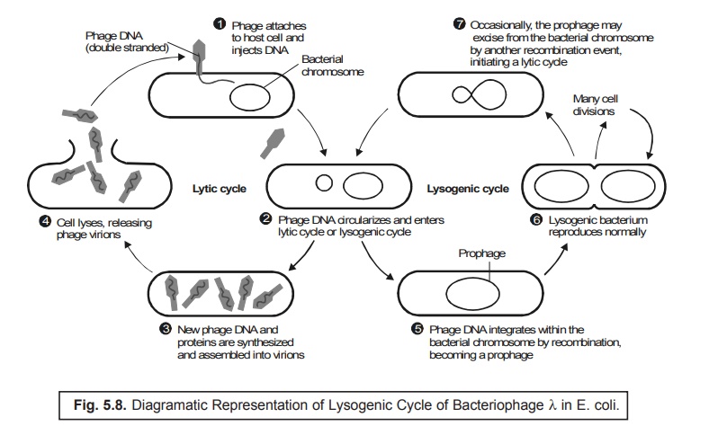
3. Bacteriophage Lambda : The Lysogenic Cycle
In a
broader and precise perspective the bacteriophage
may conveniently exist in three
phages, namely :
(a) As a free particle virion,
(b) In a lysogenic state as a prophage, and
(c) In
the vegetative state i.e., lytic cycle.
One may,
however, observe that virion is
inert in nature ; and hence, cannot reproduce. Salient Features : The various
salient features of the
bacteriophage lambda are as stated
under :
(1) In
the critical ‘lysogenic state’, the
DNA of the phage is duly integrated very much within the bacterial DNA. It usually exists in a non-infectious form known as
the prophage, and adequately replicates in synchrony with the bacterial
DNA.
(2) In
the corresponding ‘lytic cycle’, the
phage particle infects the susceptible host, undergoes multiplication, and
ultimately causes the lysis of the bacterial cell with the concomitant release
of the progeny virus particles.
(3) In a
situation when the integrated phage
is carefully induced to become the corresponding vegetative phage, the lytic
cycle comes into being.
(4) Such phages which specifically give rise to
the phenomenon of ‘lysis’ are
normally termed as the virulent phages,
as opposed to such phages that may
exist in a lysogenic state and are
usually called as the ‘temperate phages’.
(5) The
microorganisms that particularly carry the ‘temperate
phages’ are invariably termed as the ‘lysogenic
bacteria’, which are observed to be absolutely immune to the ensuing superinfection caused by the same phage.
Figure
5.8 diagramatically illustrates the lysogenic
cycle of bacteriophage λ in E. coli.
However,
it is pertinent to state here that whether decisively the ‘lytic’ or the ‘lysogenic’
response takes place immediately following infection by a temperate phage will solely depend upon both the bacterium and the phage.
Related Topics
