Microscopy : The Different Instruments
| Home | | Pharmaceutical Microbiology | | Pharmaceutical Microbiology |Chapter: Pharmaceutical Microbiology : Identification of Microorganisms
Microscopy essentially deals with the following three cardinal goals, namely : (i) Examination of ‘objects’ via the field of a microscope, (ii) Technique of determining particle size distribution by making use of a microscope, and (iii) Investigation based on the application of a microscope e.g., optical microscopy, electron microscopy.
Microscopy
: The Different Instruments
Microscopy essentially deals with the
following three cardinal goals,
namely :
(i) Examination
of ‘objects’ via the field of a microscope,
(ii) Technique
of determining particle size distribution by making use of a microscope, and
(iii) Investigation
based on the application of a microscope e.g.,
optical microscopy, electron microscopy.
Concepts
It is
worthwhile to mention here that before one looks into the different instruments
related to microscopy one may have
to understand the various vital and important cocepts, such as :
Light microscopes usually
make use of glass lenses so as to
either bend or focus the emerging
light rays thereby producing distinct enlarged
images of tiny objects. A light
micro-scope affords resolution which
is precisly determined by two
guiding factors, namely :
(a) Numerical
aperture of the lens-system, and
(b) Wavelength
of the light it uses :
However,
the maximum acheivable resolution is approximately. 0.2 μm.
Light microscopes that are
commonly employed are : the Bright field, Darkfield, Phase-contrast and
Fluorescence microscopes. Interestingly, each different kind of these
variants give rise to a distinctive image ; and, hence may be
specially used to visualize altogether different prevailing aspects of the so
called microbial morphology.
As a
rather good segment of the microorganisms are found to be almost virtually
colour-less ; and, therefore, they are not so easily visible in the Bright field Microscope directly which
may be duly fixed and stained before any observation.
One may
selectively make use of either simple or
differential staining (see Section 4.6.1.1.) to spot and visualize such
particular bacterial structures as : capsules,
endospores and flagella.
The Transmission Electron Microscope
accomplishes real fabulous resolution (approx. 0.5 nm) by employing direct electron beams having very short
wave length in comparison to the visible light.
The Scanning Electron Microscope may used
to observe the specific external
features quite explicitely, that produces an image by meticulously scanning
a fine electron beam onto the surface of specimens directly in comparison to
the projection of electrons through them.
Advent of
recent advances in research has introduced two
altogether newer versions of microscopy thereby
making a quantam jump in the improvement and ability to study the microor-ganisms and molecules in greater depth, such as :
(a) Scanning Probe Microscope ; and (b) Scan-ning Laser
Microscope.
Microscope Variants
Microbiology invariably deals with a host of
microorganisms which are practically invisible with the unaided eye. This particular discipline essentially
justifies the evolution of a variety of
micro-scopes with crucial importance so that the scientists could carry out
an elaborated and meaningful research.
The
variants in microscopes are as
stated under :
(a) Bright-field
Microscope,
(b) Dark-field
Microscope,
(c) Phase-contrast
Microscope,
(d) Differential
Interference Contrast (DIC) Microscope,
(e) Fluorescence
Microscope, and
(f) Electron
Microscope.
The
aforesaid microscope variants shall
now be treated individually and briefly in the sections that follows.
(a) Bright-Field Microscope
In
actual, practice, the ‘ordinary
microscope’ is usually refereed to as a bright-field micro-scope by virtue of the fact that it gives rise
to a distinct dark image against a brighter background.
Description : Bright-field microscope essentially
comprises of a strong metalic body with a
base and an arm to which the various other components are duly attached as
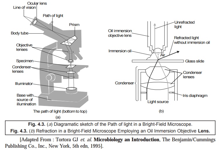
shown in Fig. 4.3 (a). It is provided
with a ‘light source’ either an
electric bulb (illuminator) or a plano-concave mirror strategi cally positioned
at the base. Focusing is accomplished by two
knobs, first, coarse adjustment knob, and secondly,
fine adjustment knob which are duly
located upon the arm in such a manner that it may move either the nosepiece or the stage so as to focus the image sharply.
In fact,
the upper segment of the microscope rightly holds the body assembly to which is
attached a nosepiece or eyepiece(s) or oculars. However, the relatively advanced microscopes do possess
eyepieces meant for both the eyes, and are legitimately termed as binocular microscopes. Importantly, the
body assembly comprises of a series of mirrors
and prisms in order that the tubular
structure very much holding the eyepiece could be tilted to afford viewing convenience. As many as 3 to 5 objectives having lenses of varying magnifying power that may be
carefully rotated to such a position which helps in clear viewing of any
objective help under the body assembly. In the right ideal perspective a
microscope must be parfocal*.
In order
to achieve high magnification (×
100) with markedly superb resolution, the lens
should be of smaller size. Though it is very desired that the light travelling via the specific specimen as well as the
medium to undergo refraction in a
different manner, at the same time it is also preferred not to have any loss of
light rays after they have gained passage via
the stained specimen. Therefore, to
preserve and maintain the direction of light rays at the maximum magnification, an immersion
oil is duly placed just between the ‘glass
slide’ and the ‘oil immersion
objective lens’, as depicted in Fig. 4.3 (b). Interestingly, both ‘glass’
and ‘immersion oil’ do possess the same refractive index ; and, therefore,
rendering the ‘oil’ as an integral
part of the optics of the glass of the microscope. In fact, the ‘oil’ exerts more or less the identical
effect as would have been accomplished by enhancing the diameter of the ‘objective’ ; and, therefore, it
critically and significantly elevates
the resolving power of the lenses. Thus, the condenser gives rise to a bright-field illumination.
(b) Dark-Field Microscope
A
Dark-Field microscope is employed particularly for the examination of ‘living
microbes’ which are either invisible in the ordinary light microscope i.e.,
cannot be properly stained by standard methods, or get distorted to a great
extent after due staining that their characteristic features may not be
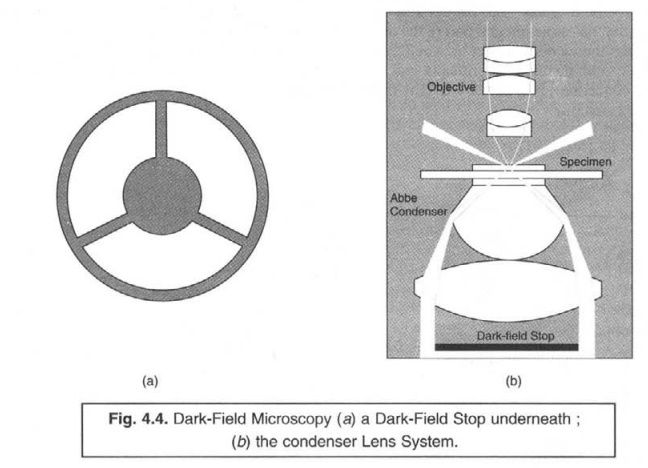
identified satisfactorily. It essentially makes use of darkfield condenser
comprising of an ‘opaque disc' instead of a normal condenser. In this
particular instance the opaque disc blocks the passage of light completely
which would have gained entry into the objective almost directly. Thus, the
only light which is specifically reflected back the specimen under examination
actually enters the objective lens precisly as illustread in Fig. 4.4(a) and
(b).
The
Dark-Field Microscopy has been successfully used in the following highly
specific investi¬gative studies, such as :
(a) Examination
of unstained bacteria suspended in an appropriate liquid,
(b) Studies
related to the internal structure as observed in eukaryotic microbes, and
(c) Examination
of highly specific and very thin spirochaetes e.g., Treponema pallidum - the
causative agent of syphilis.
(c) Phase-Contrast Microscope
The
development of the phase-contrast microscope came into being on account of the
fact that generally the unpigmented living cells fail to show their presence
vividly in the briglit-field micro¬scope as there exists practically no
difference in contrast between the cells and water. Therefore, it became almost
necessary to have the microorganisms first fixed and stained before carrying on
with the observational procedures in order to enhance the much desired contrast
as well as to produce distinct variations in colour composition between the
prevailing cell structures.
Importantly,
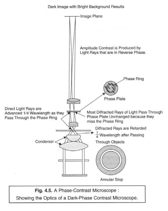
a phase-contrast microscope helps in the conversion of ensuing minimal
differ¬ences both in the refractive index and cell density directly into appreciable
detectable variations in the ‘light intensity’ ; and. therefore, ultimately
serves as an excellent means and device to observe the living cells most
conveniently, as shown in Fig. 4.5.
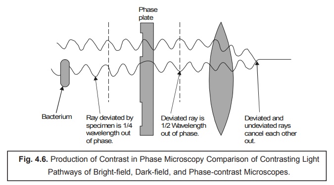
The
actual behaviour of both undeviated and deviated or even undiffracted rays in
the dark- phasc-contrast microscope is depicted in Fig. 4.6. As the light rays
do have a tendency to cancel each other out significantly, the final observed
image of the specific specimen under investigation shall ap¬pear as dark
against a relatively brighter background.
The
ensuing contrast-light pathways of bright-field, dark-field, and phase-contrast
microscopes have been explicitely illustrated in the following Fig. 4.7.
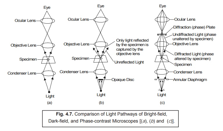
(a) Bright-field : Shows
the path of light in the bright-field
microscopy i.e., the specific
kind of illumi-nation produced by regular compound light microscopes.
(b) Dark-field : Depicts the path of light in the
dark-field microscopy i.e., it makes use of a special
condenser having an opaque disc which categorically discards all light rays in
the very centre of the beam. Thus, the only light which ultimately reaches the
specimen is always at an angle ; and thereby the only light rays duly reflected
by the specimen (viz., gold rays) finally reaches the objective lens.
(c) Phase-contrast :
Illustrates the path of light in the phase-contrast
microscopy i.e., the light rays
are mostly difracted altogether in a different manner ; and, therefore, do
travel various pathways to reach the eye of the viewer. Thus, the diffracted light rays are duly
indicated in gold ; whereas, the undiffracted light rays are duly shown
in red.
(d) Differential Interference Contrast (DIC) Microscope
The differential interference contrast (DIC
microscope bears a close resemblance to the phase-contrast microscope (Section 4.6.2.2.3.) wherein it
specifically produces an image based upon the ensuing differences in two
fundamental physical parameters, namely : (a)
refractive indices ; and (b) thickness.
In acutal practice, two distinct and prominent beams of
plane-polarized light strategically held at right angles to each other are duly
produced by means of prisms. Thus, in one of the particular set-ups, first the object beam
happens to pass via the specimen ; and secondly the reference beam
is made to pass via a clear zone in the slide. Ultimately,
after having passed via the
particular specimen, the two emerging
beams are combined meticulously thereby causing actual interference with each other to give rise to the formation
of an ‘image’.
Applications : DIC-microscope helps to
determine :
(1) Live,
unstained specimen appears usually as 3D highly coloured images.
(2) Clear
and distinct visibility of such structures : cell walls, granules, vacuoles,
eukaryotic nuclei, and endospores.
Note : The resolution of a DIC-Microscope is
significantly higher in comparison to a standard phase-contrast microscope, due
to addage of contrasting colours to the specific specimen.
(e) Fluorescence Microscope
Interestingly,
the various types of microscopes
discussed so far pertinently give rise to an ‘im-age’ from light which happens to pass via a specimen. It is,
however, important to state here that the
fluorescence microscopy exclusively based upon the inherent fluorescence characteristic feature of a ‘substance’
i.e., the ability of an object
(substance) to emit light distinctly. One may put forward a plausible
explanation for such an unique physical phenomenon due to the fact that ‘certain molecules’ do absorb radiant* energy thereby
rendering them highly excited ; however, at a later convenient stage
strategically release a reasonable proportion of their acquired trapped energy
in the form of ‘light’ (an energy).
It has been duly proved and established that any light given out by an excited molecule shall possess definitely a longer wavelength (i.e., having lower energy) in comparison to the radition absorbed
initially’.
Salient Features : There are
certain ‘salient features’ with
regard to the fluorescence microscopy as
stated under :
(1) Quite
a few microbes do fluoresce naturally on being subjected to ‘special lighting’.
(2) Fluorochromes : In such
an instance when the ‘specimen under
investigation’ fails to fluoresce
normally, it may be stained adequately with one of a group of fluorescent dyes
termed as ‘fluorochromes’.
(3) Microorganisms upon
staining with a fluorochrome when examined with the help of a fluorescence microscope in an UV or near-UV light source, they may
be observed as luminescence bright
objects against a distinct dark background.
Examples : A few typical examples are as
follows :
(a) Mycobacterium
tuberculosis : Auramine O
(i.e., a fluorochrome) that usually
glows yel-low on being exposed to
UV-light, gets strongly absorbed by M. tuberculosis (a pathogenic
‘tuberculo-sis’ causing organism). Therefore, the dye when applied to a specific sample being investigated for
this bacterium, its presence may be
detected by the distinct visualization of bright yellow microbes against a dark
background,
(b) Bacillus
anthracis : Fluorescein
isothiocyanate (FITC) (i.e., a
fluorochrome) stains B. anthracis particularly and appears as ‘apple
green’ distinctly. This organism
is a causative agent of anthrax.

Fig. 4.8.
illustrates the diagramatic sketch of the vital components and the underlying
principles of operation of a Fluorescence Microscope.
Methodology : The various steps involved in the
operational procedures of a fluorescence
mi-croscope are as under :
(1) A
particular specimen is exposed to UV-light
or blue light or violet light thereby giving rise to the
formation of an image of the ‘specified
object’ along with the resulting fluorescence light.
(2) A
highly intense beam is duly generated
either by a Mercury Vapour Lamp (a) or any other appropriate source ;
and the ensuing heat transfer is
duly limited by a specially designed Infra-red
Filter (b).
(3) Subsequently,
the emerged light is made to pass through an Exciter Filter (c) which
allows the specifc transmission of exclusively the desired wavelength.
(4) A Darkfield Condenser (d) critically affords a black background against which the fluo-rescent objects usually glow.
(5) Invariably
the particular specimen is stained with
fluorochrome (dye molecule) (e), which ultimately fluoresces brightly on
being exposed to light of a particular wavelength ; whereas, there are certain
organisms that are autofluorescing
in nature.
(6) A Barrier Filter (f) is strategically
positioned after the objective Lenses
(g) helps to re-move any residual UV-light thereby causing two important functional advantages,
namely :
(i) To
protect the viewer’s eyes from
getting damaged, and
(ii) To
suitably eliminate the blue and violet
light thereby minimising the image’s actual contrast.
Applications : The various useful applications
of a Fluorescence Microscope are as
enumerated under :
(1) It
serves as an essential tool in both ‘microbial
ecology’ and ‘medical microbiology’.
(2) Important
microbial pathogens like : M.
tuberculosis may be distinctly identified in two different modalities, for instance :
(i) particularly
labeling the microorganisms with fluorescent
antibodies employing highly specialized immunofluorescence techniques, and
(ii) specifically
staining them (microbes) with fluorochromes.
(3) Ecological investigative studies is
usually done by critically examining the specific micro-organisms duly stained
with either fluorochromes e.g., acridine orange, and diamidino-2-phenylindole
(DAPI)-a DNA specific stain or
fluorochrome-labeled probes.
(f) Electron Microscope
An electron microscope refers to a
microscope that makes use of streams of
electrons duly deflected from their course either by an electrostatic or by an electromagnetic
field for the magnifica-tion of objects. The final image is adequately viewed
on a fluorescent screen or recorded on a photo-graphic plate. By virtue of the
fact that an electron microscope
exhibits greater resolution, the ensuing images may be magnified conveniently
even upto the extent of 4,00,000
diameters.
It is,
however, pertinent to mention here that objects that are smaller than 0.2 μm, for
instance : internal structures of cells,
and viruses should be examined,
characterized and identified by the aid of
an electron microscope.
Importantly,
the electron microscope utilizes
only a beam of electrons rather than
a ray of light. The most acceptable, logical, and plausible explanation that
an electron microscope affords much prominent and better resolution
is solely on account of the fact that the ‘electrons’
do possess shorter wavelengths
significantly. Besides, the
wavelengths of electrons are approximately 1,00,000 times smaller in comparison to the wavelengths of visible light.
Interestingly,
an electron microscope predominantly
employs electromagnetic lenses,
rather than the conventional glass
lenses in other microscopes ; and ultimately, focused upon a ‘specimen’ a beam of electrons which is made to travel via a tube under vacuum (so as to eliminate any loss of energy due to friction collision
etc.).
Types of Electron Microscopes :
The electron microscopes are of two different types, namely :
(a) Transmission
Electron Microscope (TEM), and
(b) Scanning
Electron Microscope (SEM).
These two
types of electron microscopes shall
be discussed briefly in the sections that follows :
(a) Transmission Electron Microscope (TEM)
The transmission electron microscope (TEM)
specifically makes use of an extremely
fine focused beam of electrons released
precisely from an ‘electron gun’ that
penetrates via a specially prepared ultrathin section of the investigative specimen, as illustrated in
Fig. 4.9.
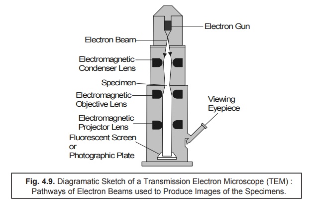
Methodology : The various operational steps and
vital components of TEM are described below :
(1) The Electron Gun i.e., a pre-heated Tungsten
Filament, usually serves as a beam
of elec-trons which is subsequently focussed upon the desired ‘specimen’ by the help of Electromagnetic Condeser Lens.
(2) Because
the electrons are unable to
penetrate via a glass lens, the usage
of doughnut-shaped electromagnets usually termed as Electromagnetic Objective Lenses are made so as to focus the beam
properly.
(3) The
entire length of the column comprising of the vairous lenses as well as
specimen should be maintained under high
vacuum*.
(4) The specimen causes the scattering of electrons that are
eventually gaining an entry through it.
(5) The ‘electron beam’ thus emerged is
adequately focused by the aid of Electromagnetic
Projector Lenses strategically
positioned which ultimately forms an
enlarged and distinctly visible
image of the ‘specimen’ upon a Fluorescent Screen (or Photographic Plate).
(6) Specifically
the appearance of a relatively denser
region in the ‘specimen’ helps
to scatter much more electrons ; and, hence, may be viewed as darker zones in the image because only
fewer electrons happen to touch that particular zone of the fluorescent screen (or photographic plate).
(7) Finally,
in a particular contrast situation,
the electron-transparent zones are
definitely brighter always. The ‘screen’
may be removed and the image may be captured onto a ‘photographic plate’ to
obtain a permanent impression as a record.
(b) Scanning Electron Microscope (SEM)
As it has
been discussed under TEM that an image can be obtained from such
radiation which has duly transmitted through a specimen. In a most recent technological advancement the Scanning Electron Microscope (SEM) has been developed whereby detailed
in-depth examination of the sur-faces of
various microorganisms can be accomplished with excellent ease and
efficiency. In reality, the SEM markedly differs from several other
electron microscopes wherein the
image is duly obtained right from the electrons that are strategically emitted
by the surface of an object in
comparison to the transmitted electrons.
Thus, there are quite a few SEMs which
distinctly exhibit a resolution of 7 nm or
even less.
Fig. 4.10. duly depicts the diagramatic sketch fo a Scanning Electron Microscope (SEM) that vividly shows the primary electrons sweeping across the particular investigative specimen together with the knock electrons emerging from its surface. In actual practice, these secondary electrons (or knock electrons) are meticulously picked up by a strategically positioned collector, duly amplified, and transmitted onto a viewing screen or a photographic plate (to have a permanent record/impression of the investigative specimen).
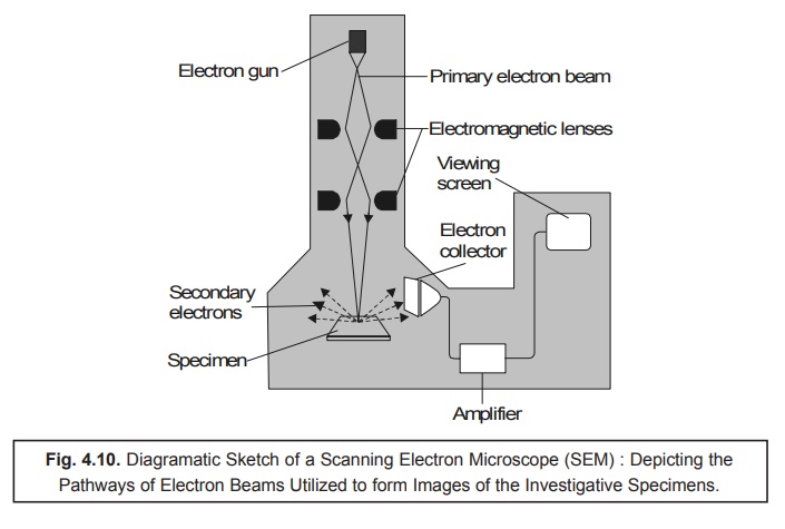
Methodology : The various steps involved in the
operative sequential steps are as stated under :
(1) Specimen preparation : It is
quite simple and not so cumbersome ; and even in certain cases one may use air-dried specimen for routine examination
directly. In general, largely the microor-ganisms should first be fixed,
dehydrated, and dried meticulously so as to preserve
not only the so called ‘surface-structure’
of the specimen but also to prevent the ‘possible collapse of
the cells’ when these are
directly exposed to the SEM’s high
vacuum. Before, carrying out the usual viewing activities, the dried samples
are duly mounted and carefully coated with a very thin layer of metal sheet in
order to check and prevent the buildup of an accumulated electrical charge onto
the surface of the specimen and to provide a distinct better image.
(2) SEM helps in the scanning of a
relatively narrow and tapered electron beam both back and forth onto the surface of the specimen. Thus, when the beam of
electron happens to strike a specific zone of the specimen, the surface atoms
critically discharge a small shower of electrons usually termed as ‘secondary electrons’, which are
subsequently trapped and duly registered by a specially designed detector.
(3) The ‘secondary electrons’ after gaining
entry into the ‘detector’ precisely
strike a scintillator thereby enabling
it to emit light flashes which is
adequately converted into a stream of elctrical current by the aid of a photomultiplier tube. Finally, the
emerging feeble electrical current is duly amplified.
(4) The
resulting signal is carefully sent across to a strategically located cathode-ray tube, and forms a sharp
image just like a television picture, that may be either viewed or photographed
accord-ingly for record.
Notes :
(i) The actual and exact number of secondary
electrons that ultimately reach the ‘detector’ exclu-sively depends upon the
specific nature of the surface of the investigative specimen.
(ii) The ensuing ‘electron beam’ when strikes a
raised surface area, a sizable large number ‘sec-ondary electrons’ gain due
entry into the ‘detector’ ; whereas, fewer electrons do escape a depression in
the surface of the specimen and then reach the detector. Therefore, the raised
zones appear comparatively lighter on the screen, and the depressions are
darker in appear-ance. Thus, one may obtain a realistic 3D image of the surface
of the microorganism having a visible intensive depth of focus.
Related Topics

