Adipose Tissue In Fasting
| Home | | Biochemistry |Chapter: Biochemistry : The Feed-Fast Cycle
Glucose transport by insulin-sensitive GLUT-4 into the adipocyte and its subsequent metabolism are depressed due to low levels of circulating insulin. This results in decreased TAG synthesis.
ADIPOSE TISSUE IN FASTING
A. Carbohydrate metabolism
Glucose transport by
insulin-sensitive GLUT-4 into the adipocyte and its subsequent metabolism are
depressed due to low levels of circulating insulin. This results in decreased
TAG synthesis.
B. Fat metabolism
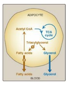
1. Increased degradation of fat: The PKA-mediated phosphorylation
and activation of HSL and subsequent hydrolysis of stored fat are enhanced by
the elevated catecholamines norepinephrine and epinephrine. These hormones,
which are released from the sympathetic nerve endings in adipose tissue and/or
from the adrenal medulla, are physiologically important activators of HSL
(Figure 24.14,1).
2. Increased release of fatty acids: FAs obtained from hydrolysis of
stored TAG are primarily released into the blood (see Figure 24.14, 2). Bound to albumin, they are
transported to a variety of tissues for use as fuel. The glycerol produced from
TAG degradation is used as a gluconeogenic precursor by the liver, which
contains glycerol kinase. [Note: FA can also be oxidized to acetyl CoA, which
can enter the TCA cycle, thereby producing energy for the adipocyte. They also
can be re-esterified to glycerol 3-phosphate (from glyceroneogenesis,),
generating TAG and reducing plasma FA concentration.]
3. Decreased uptake of fatty acids: In fasting, LPL activity of
adipose tissue is low. Consequently, circulating TAG of lipoproteins is not
available to adipose tissue.

Figure 24.14 Major metabolic
pathways in adipose tissue during fasting. [Note: The numbers in the circles,
which appear both in the figure and in the corresponding citation in the text,
indicate important pathways for fat metabolism.] CoA = coenzyme A; TCA =
tricarboxylic acid.
Related Topics
