Bacteria [Plural of ‘Bacterium’]
| Home | | Pharmaceutical Microbiology | | Pharmaceutical Microbiology |Chapter: Pharmaceutical Microbiology : Characterization, Classification and Taxonomy of Microbes
Exhaustive historical evidences based on the survey of literatures, amply stress and reveal the fact that ‘bacteria’ predominantly share with the ‘blue-green algae’ a unique status and place, in the ‘world of living organisms’.
Bacteria
[Plural of ‘Bacterium’]
Exhaustive
historical evidences based on the survey of literatures, amply stress and
reveal the fact that ‘bacteria’
predominantly share with the ‘blue-green
algae’ a unique status and place, in the ‘world of living organisms’.
A bacterium may be regarded as a
one-celled organism without true nucleus or functionally specific components of metabolism that essentially belongs to the
kingdom Prokaryotae (Monera), a name
which means primitive nucleus. However, all other living organisms are termed
as Eukaryotes, a name that precisely
implies a true or proper nucleus.
It has
been duly observed and established that ‘bacteria’
are exclusively responsible for the causation of several painful ailments in
humans, namely : tonsillitis, pneumonia,
cystitis, school sores, and conjunctivitis.
Alternatively,
one may define bacteria as
microscopic single-celled organisms that can pen-etrate into healthy tissues
and start multiplying into vast numbers. Interestingly, when they do this, they
invariably damage the tissue that they are infecting, causing it to break down
into the formation of pus.** Due to
the damage they (bacteria) cause, the affected and involved area becomes red,
swollen, hot and painful. In this
manner, the waste products of the damaged tissue, together with the bacteria,
rapidly spread into the blood stream, and this virtually stimulates the brain
to elevate the body tempera-ture so as to fight off the contracted infection; and
this ultimately gives rise to the development of ‘fever’ (normal body temperature being 37°C or 98.4°F).
Salient Features
The
various salient features of ‘bacteria’ are as stated under:
(1) The
body is invariably invaded by millions of organisms every day, but very few
surprisingly may ever succeed in causing serious problems by virtue of the fact
that body’s defence mechanisms usually destroy the majority
of the invading microbes.
In fact,
the white-blood cells (WBCs) are the main line of defence against the
prevailing infections. Evidently, the WBCs rapidly migrate to the zone of
‘unwanted bacteria’ and do help in engulfing them and destroying them
ultimately. Importantly, when these defence mechanisms get overwhelmed, that a
specific infection develops and noticed subsequently.
(2) Nomenclature: Each
species of organisms or bacteria (and fungi but not viruses) has two
names: first — a family name (e.g., Staphylococcus)
that essentially makes use of a capital initial
letter and comes first always; and secondly
— a specific species name (e.g., aureus) which uses a lower case initial
letter and comes second.
Example: The golden staph bacteria that
gives rise to several serious throat infections is therefore termed as Staphylococcus
aureus, but should be normally abbreviated to S. aureus.
(3) As
different types of bacteria invariably favour different segments of the body
and thereby lead to various glaring symptoms; therefore, it is absolutely
necessary to choose and pick-up an appropriate educated guess about the
antibiotic(s) to be administered by a ‘physician’.
In the event of any possible doubt it is always advisable to take either a ‘sample’ or a ‘swab’ being sent to a ‘microbiological
laboratory’ for an expert analysis, so that the precise organ-ism may be identified, together with the most
suitable antibiotic to destroy it completely.
(4) Obviously,
there are a plethora of organisms (bacteria), specifically those present in the
‘gut’, observed to be quite useful
with respect to the normal functioning of the body. These organisms usually help in the digestive process, and prevent
infections either caused by fungi (e.g., thrush) or sometimes by viruses.
Importantly, antibiotics are capable
of killing these so called ‘good bacteria’ also, which may ultimately
give rise to certain apparent side-effects due to the prolonged usage of
antibiotics, such as : diarrhoea, fungal
infections of the mouth and vagina.
The most
commonly observed ‘bacteria’ that
invariably attack the humans and the respective diseases they cause, or organs
they attack, are as listed under :
Bacteria : Diseases or Place of Infections
Bacteroides
: Pelvic organs
Bordetella
pertussis : Whooping cough
Brucella
abortus : Brucellosis
Chlamydia
trachomatis : Vineral disease, pelvic organs, eye
Clostridium
perfringens : Gas gangrene, pseudomembranous colitis.
Clostridium
tetani : Tetanus
Corynebacterium
diphtheriae : Diphtheria
Escherichia
coli : Urine, gut, fallopian tubes, peritonitis
Haemophilus
influenzae : Ear, meningitis
Helicobacter
pylori : Peptic ulcers
Klebsiella
pneumoniae : Lungs, urine
Legionella
pneumophilia : Lungs
Mycobacterium
leprae : Leprosy
Mycobacterium
tuberculosis : Tuberculosis
Mycoplasma
pneumoniae : Lungs
Neisseria
gonorrhoea : Gonorrhoea, pelvic organs
Proteus :
Urine, ear
Pseudomonas
aeruginosa : Urine, ear, lungs, heart
Salmonella
typhi : Typhoid
Shigella
dysenteriae : Gut infections
Staphylococcus
aureus : Lungs, throat, sinusitis, ear, skin, eye, gut, meningitis, heart,
bone, joints
Streptococcus
pyrogens : Sinuses, ear, throat, skin
Streptococcus
viridans : Heart
Treponema
pallidum : Syphillis
Yersinia
pestis : Plague
Structure and Form of the Bacterial Cell:
These
characteristic form of the bacterial cell may be sub-divided into two
heads, namely:
(i) Size
and shape, and
(ii) Structure
These two categories shall now be dealt with
separately in the sections that follows :
Size and Shape:
The size and shape of bacteria largely vary between the dimensions of
0.75 - 4.0 μm. They
are invariably obtained as definite
unicellular structures that may be essentially found either as spherical forms (i.e., coccoid forms) or
as cylindrical forms (i.e., rod-shaped forms). How-ever, the latter forms, in one or two
genera, may be further modified into two
sub-divisions, namely:
(a) With
a single twist (or vibrios), and
(b) With several twists very much akin to ‘cork screw’ (or spirochaetes).
In actual
practice, there prevails another predominant characteristic feature of the
bacterial form i.e., the inherent
tendency of the coccoid cells to exhibit
growth in aggregates. It has been duly observed that these ‘assemblies’
do exist in four distinct manners,
such as:
(i) As ‘pairs’ (or diplococci),
(ii) As ‘groups of four systematically arranged in a
cube’ (or sarcinae),
(iii) As ‘unorganized array like a bunch of grapes’
(or staphylococci), and
(iv) As ‘chains like a string of beads’ (or streptococci).
In
general, these ‘aggregates’ are so
specific and also characteristic that they usually assign a particular generic
nomenclature to the group, for instance :
(a) Diplococcus pneumoniae — causes
pneumonia,
(b) Staphylococcus aureus — causes
‘food-poisoning’ and boils, and
(c) Streptococcus
pyogenes — causes severe sore throat.
Structure:
There
exists three essential divisions of
the so called ‘bacterial cell’ that
normally occur in all species, such as: cell
wall or cytoplasmic membrane and cytoplasm.
Based
upon the broad and extensive chemical investigations have evidently revealed two funda-mental components in the
structure of a bacterial cell, namely :
(a) Presence
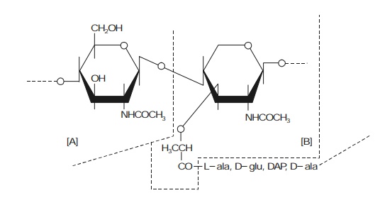
of a basic structure of alternating N-acetyl-glucosamine,
and
(b) N-acetyl-3-0-1-carboxyethylglucosamine
molecules. In fact, the strategic union of the said two components distinctly give rise to the polysaccharide backbone.
Salient Features: The salient features of the structure of a
bacterial cell are as stated under:
(1) The
two prominent and identified chemical entities viz., N-acetyl glucosamine (A), and N-acetyl-3-0-1-
carboxymethylglucosamine (B) are usually cross-linked by peptide chains as
shown under:

(2) The
combined structure of [A] and [B] as shown in (1) above basically possesses an enor-mous mechanical strength, and,
therefore, essentially represents the target for a specific group of ‘antibiotics’,
which in turn via different modes,
categorically inhibit the biosynthesis
that eventually take place either in the course of cell growth or in the cell
division promi-nently.
(3) The
fundamental peptidoglycan moiety
(also known as murein or mucopeptide) besides contains other
chemical structures that particularly gets distinguished by the presence of two kinds of bacteria, namely :
(a) Gram-negative
organism, and
(b) Gram-positive
organisms.
However,
these two variants of organisms may
be identified distinctly and easily by treating a thin-film of bacteria, duly
dried upon a microscopic slide with a separately prepared solution of a basic dye i.e., gentian violet, and followed soon shape after by the
application of a solution of iodine. Thus, we may have:
Gram-negative bacteria — by
alcohol washing the dye-complex from certain types of cells, and Gram-positive bacteria — by retaining the dye-complex despite the prescribed
alcohol-washing.
Further,
the prevailing marked and pronounced differences in behaviour, just discovered
by a stroke of luck, are now specifically recognized to be a glaring reflection
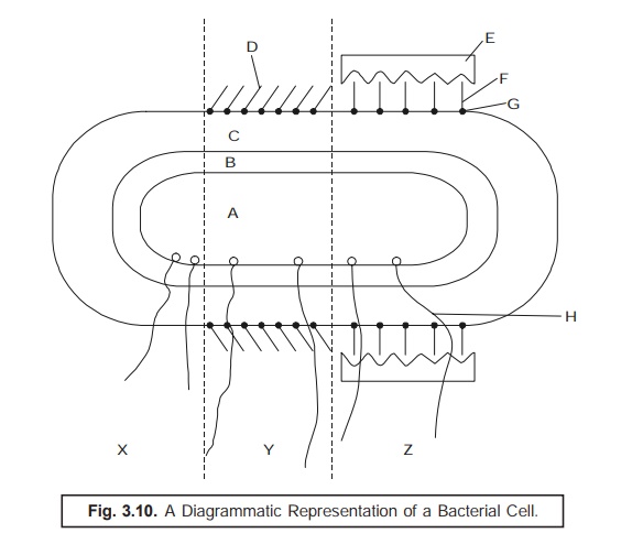
of wall structure variants in the two kinds of cell as illustrated in Fig.
3.10.

X =
Generalized structure of a Bacterial Cell;
Y = Gram
+ ve Structure;
Z = Gram
– ve Structure
A =
Cytoplasm;
E =
Lipopolysaccharide;
B =
Cytoplasm membrane;
F =
Lipoprotein;
C =
Cell-Wall peptidoglycan;
G =
Covalent-Bond;
D =
Teichoic acid;
H =
Flagellum;
Gram-positive Cell Wall [Y] : In this
particular instance, the walls of bacteria essentially com-prise of the
molecules of a polyribitol or polyglycerophosphate that are found to
be strategically attached by means of covalent
bonds (G) to the prevailing oligosaccharide
backbone; and these chemical entities are nothing but teichoic acids [D]. It is, however, pertinent to mention here, that
the said teichoic acids do not give rise to any sort of
additional rigidity upon the ensuing cell wall, but as they are acidic in
character, they are capable of sequestering essential metal cations derived
from the culture media upon which the
bacterial cells are growing. Importantly, this could be of immense value in
such circum-stances wherein the ‘cation
concentration’ in the environment is apparently at a low ebb.
Gram-negative Cell Wall : Interestingly,
the Gram-negative cell wall is
observed to be much more complex in
character by virtue of the presence of the lipoprotein molecules (F)
strategically at-tached covalently to the respective vital oligosaccharide
backbone. Besides, on its outer region, a layer of lipopolysaccharide (E) along
with the presence of protein critically attached by hydrophobic interac-tions
and divalent metal cations e.g., Ca2+,
Fe2+, Mg2+, Cu2+, whereas, in its inner side
is a layer of phospholipid.
Related Topics
