Adrenal Glands
| Home | | Anatomy and Physiology | | Anatomy and Physiology Health Education (APHE) |Chapter: Anatomy and Physiology for Health Professionals: Endocrine System
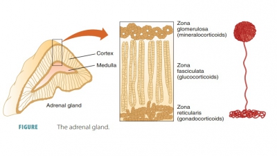
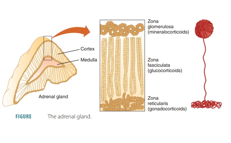
The pyramid-shaped adrenal glands sit on top of each kidney like caps and are embedded in the adipose tis-sue enclosing each kidney.
Adrenal
Glands
The pyramid-shaped adrenal
glands sit on top of each kidney like caps and are embedded in the adipose
tissue enclosing each kidney (FIGURE
16-15). The adre-nal glands are also known as the suprarenal glands because of their position.
The adrenal glands have a central adrenalmedulla and an outer adrenal cortex, each secret-ing
different hormones. The adrenal medulla is closely connected with the
sympathetic division of the autonomic nervous system and appears more like a
mass of nervous tissue than a gland. The adrenal cortex consists of layers of
cells, including an outer zone or zona
glomerulosa, a middle zone or zonafasciculate
, and an inner zone or zona
reticularis. Itis derived from the embryonic mesoderm. Both the adrenal medulla and cortex are well supplied with blood
vessels.

Adrenal Cortex
The adrenal cortex synthesizes more than 30 corticosteroids. This synthesis begins with
choles-terol, using a variety of intermediate substances in relation to the
hormone being made. Steroid hor-mones are not stored in cells and their rate of
release is based on their rate of synthesis. Some of these hormones are vital
for survival, especially aldoste-rone, cortisol, and some sex hormones.
Mineralocorticoids
Mineralocorticoids
mainly function in the regula-tion of mineral salt or electrolyte
concentrations in the extracellular fluids, mostly regulating sodium and
potassium. The regulation of sodium is connected to the regulation of
potassium, hydrogen, bicarbonate, and chloride. The regulation by
mineralocorticoids of sodium and potassium is vital for overall homeo-stasis.
Aldosterone accounts for more than 95% of the mineralocorticoids produced.
The outer adrenal cortex or zona glomerulosa synthesizes aldosterone, a mineralocorticoid that helps regulate mineral electrolyte concentrations. Aldosterone helps the kidneys to balance sodium and potassium and stimulates water retention via the process of osmosis. If blood sodium decreases or blood potassium increases, the adrenal cortex secretes aldosterone. The kidneys also stimulate aldosterone secretion if blood pressure falls. Aldosterone also enhances the absorption of sodium from gastric juice, perspiration, and saliva. It has regulatory effects that occur within 20 minutes, allowing precise control of plasma electrolyte balance. The activity of aldosterone involves synthesis and activation of proteins needed for sodium transport. Aldosterone secretion is stimulated by decreased blood volume and pressure and raised blood levels of potassium. Its secretion is inhibited by the opposite conditions. The two most important mechanisms that regulate aldosterone secretion are the renin–angiotensin–aldosterone mechanism and the plasma concentrations of potassium.
The renin–angiotensin–aldosterone mechanism regulates
aldosterone release, helping to control blood volume, blood pressure, and the
reabsorption of sodium and water by the kidneys. When blood pressure or vol-ume
falls, certain cells of the juxtaglomerular
complex of the kidneys are excited, which respond by releas-ing renin into the blood.
The renin cleaves off part of the plasma protein known as angiotensinogen. This causes an enzymatic cascade
to occur forming angiotensin
II. This substance stimulates cells of
theglomerulosa to release aldosterone. All the effects of this mechanism
ultimately raise blood pressure.
The cells of the zona glomerulosa in the adrenal cortex are
directly influenced by fluctuating blood levels of potassium. When increased,
potassium stimu-lates aldosterone release, and the opposite is also true. ACTH
normally has very little effect on aldosterone release. However, when stressors
are prevalent, the hypothalamus secretes more CRH. ACTH rises in the blood,
increasing aldosterone secretion slightly. This helps to deliver nutrients and
respiratory gases in an attempt to cope with the stressors.
Atrial
natriuretic peptide is a hormone fromthe heart that is secreted when
blood pressure rises. It regulates blood pressure and sodium–water balance and
greatly inhibits the renin–angiotensin–aldosterone mechanism. Renin and
aldosterone secretion are blocked, and atrial natriuretic peptide also inhibits
other mechanisms that enhance sodium and water reabsorp-tion. Overall, it
decreases blood pressure by allowing sodium and water to leave the body in the
urine.
Glucocorticoids
In general, the glucocorticoids help us
to resist stressors and influence energy metabolism. Cortisol is a glucocorticoid produced in the
middle adrenal cortex or zona
fasiculata that also influences protein and fat metabolism. Cortisol
inhibits protein synthe-sis, promotes the conversion of lipids to glucose, and
the formation of glycogen in the liver. Cortisol, which is also known as hydrocortisone, helps balance blood
glucose and is controlled by negative feedback. ACTH stimulates the adrenal
cortex to release cortisol, and stress plays an important part in triggering
cortisol release. FIGURE
16-16 shows how negative feedback regulates cortisol secretion.
Other glucocorticoid hor-mones include cortisone
and corticosterone, but these are
relatively insignificant compared with cortisol. Acute stress interrupts normal
cortisol rhythm, result-ing in increased ACTH blood levels. Cortisol responds
to stress by causing a large rise in blood glucose, amino acids, and fatty
acids. Its metabolic effect known as glu-coneogenesis
is defined as the formation of glucose fromfats and proteins. Cortisol, in
an attempt to conserve glucose for the brain, mobilizes fatty acids from
adi-pose tissue so they can be used for energy. Stored pro-teins are broken
down, vasoconstriction is enhanced, and nutrients are dispersed to the cells
more quickly than normal. Excessive cortisol, however, causes anti-inflammatory
and anti-immune effects to a large degree. When excessive, cortisol depresses
cartilage formation, bone formation, and the immune system. It disrupts normal
cardiovascular, gastrointestinal, and neural function and inhibits inflammation
via the decrease in the release of inflammatory chemicals.

Gonadocorticoids
The inner adrenal cortex or zona reticularis pro-duces sex hormones. Under
stimulation by ACTH, this part of the adrenal cortex produces smallamounts of androgens, the sex hormones produced in
larger quantities by the testes in males. Some androgens from the zona
reticularis are converted to estrogens, which are the dominant sex hormones in
females. Adrenal androgens stimulated development of pubic hair in both sexes
prior to puberty. Adrenal sex hormones are also called gonadocorticoids. Their release is linked to ACTH. TABLE 16-5 discusses the adrenal cortex
hormones.

Adrenal Medulla
The adrenal medulla contains large, round cells that are
similar to the cells of the sympathetic ganglia, which are innervated by
preganglionic sympa-thetic fibers. The secretory activities of the adrenal
medulla are controlled by the sympathetic divi-sion of the autonomic nervous
system. The adre-nal medulla secretes epinephrine or adrenaline and
norepinephrine or noradrenaline . Epineph-rine makes up
80% of adrenal medulla secretions, the rest being norepinephrine. These
hormones aid in coping with stressors and participate in the fight-or-flight
response. Epinephrine is stronger in its stimulation of bronchial dilation and
increased blood flow to the heart and skeletal muscles. How-ever, epinephrine
decreases peristalsis. It is used clinically as a bronchodilator and heart
stimulant, since it increases heart rate and blood pressure. Nor-epinephrine
more greatly influences blood pressure and peripheral vasoconstriction.
One group of chemicals produced in nervous tissue, called catecholamines, regulate many dif-ferent
functions, including thought processes, hor-mone secretions, blood pressure,
and heart rate. The most common catecholamines are epineph-rine, norepinephrine,
dopamine, and serotonin. The adrenal medulla is a modified sympathetic
ganglion.
The adrenal medulla’s secretions have long-lasting effects.
They increase heart rate, cardiac muscle con-traction force, breathing rate,
and blood glucose level while elevating blood pressure and decreasing
diges-tive activity. In response to stress, the hypothalamus releases impulses
to control the adrenal medulla. These impulses prepare the body for the “fight-or-flight
response.” TABLE
16-6 discusses adrenal medullary hormones and their effects.

1. What are the names of the hormones released from the
adrenal glands?
2. Explain the effects of aldosterone in the kidneys.
3. Which pituitary hormone stimulates the adrenal cortex?
4. What are the three zonae of the adrenal cortex?