Pineal Gland
| Home | | Anatomy and Physiology | | Anatomy and Physiology Health Education (APHE) |Chapter: Anatomy and Physiology for Health Professionals: Endocrine System
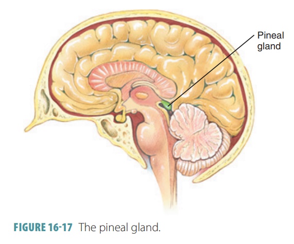
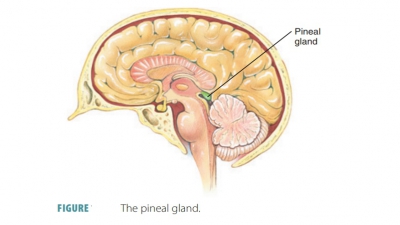
The small, cone-shaped pineal gland is located deep in the cerebral hemispheres and is attached to the thalamus near the upper part of the third ventricle.

Pineal
Gland
The small, cone-shaped pineal gland is located deep in the cerebral hemispheres and is attached to the thalamus near the upper part of the third ventricle (FIGURE 16-17). It secretes the hormone melatonin in response to daylight conditions in the external envi-ronment from its pinealocytes, which are arranged in tight cords and clusters. In adults, between the pinealocytes, calcium salts are contained in dense par-ticles. The calcium salts are radiopaque, and therefore the pineal gland is used as an aid in determining the orientation of the brain when X-rays are taken. When it is dark, nerve impulses from the eyes decrease and the secretion of melatonin increases. Melatonin is an amine hormone derived from serotonin.
Melatonin functions as a biologic clock and can help to
regulate the circadian
rhythms, which are associ-ated with environmental day and night
cycles and help the body to distinguish day from night. Melatonin is not fully
understood, but appears to inhibit gonadotropin secretion to help regulate the
female reproductive cycle and to control the onset of puberty. In children,
mela-tonin may inhibit early sexual maturation. The biologicclock of humans is the
suprachiasmatic nucleus of thehypothalamus. Its large amount of melatonin
receptors react to bright light, altering the biologic clock. There-fore,
altered melatonin levels may result in variations of appetite, body
temperature, and sleep habits.