Lever Systems
| Home | | Anatomy and Physiology | | Anatomy and Physiology Health Education (APHE) |Chapter: Anatomy and Physiology for Health Professionals: Support and Movement: Muscular System
Most skeletal muscles use leverage in order to move. To understand further, a lever is any rigid structure that moves on a fixed point, which is known as a fulcrum, when force is applied.
Lever Systems
Most skeletal muscles use
leverage in order to move. To understand further, a lever is any rigid structure that moves on a fixed point, which is known as
a fulcrum, when force is applied. This applied force is known as effort, which is used to move a load, which is a
resistance. The body’s joints act as fulcrums, while the bones act as levers. Muscle con-traction
creates the effort applied at an insertion point on a bone. The bone itself as
well as its overlying tissues and anything else being moved together act as
the load.
Levers
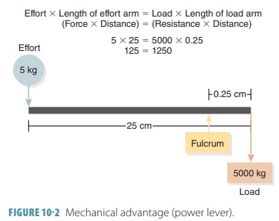
The concept of levers involves the movement of loads that are heavier, or the movement of loads at a faster speed or over a further distance. When a load is close to a fulcrum and effort is applied further away from the fulcrum, just a small effort over a relatively large distance can move a large load over a small dis-tance (FIGURE 10-2). This operation or mechanical advantage is often referred to as a power lever. Think of a car jack, in which a heavy vehicle is moved upward for only a small distance, in order to change a tire. The muscle effort needed to make the jack work is very small.
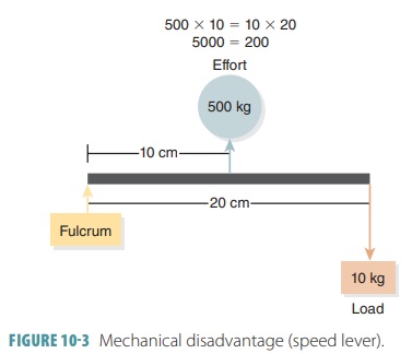
However, when a load is far from a fulcrum and effort is applied near the fulcrum, the exerted force must be greater than the load that will be supported or moved. This operation or mechanical disadvantage is often referred to as a speed lever (FIGURE 10 -3). They allow a load to be moved quickly, over a large distance, with a wide range of motion such as using a shovel to dig a hole. Small differences in the muscle’s insertion site compared to the joint can create large differences in the amount of force needed to move a certain load or resistance.
Classes of Levers
There are three basic classes of
levers, which can be understood by their comparison to certain muscle actions
in the body:
■■ First-class levers: Effort is
applied at one end while the load is at the other end and the fulcrum is
somewhere in between such as in scissors or seesaws. In the body, first-class
leverage occurs when the head is lifted off the chest. Some first-class levers
operate for strength (mechanical advantage) and some operate for speed and
distance (mechanical disadvantage).
■■ Second-class levers: Effort
is applied at one end of a lever while the fulcrum is at the other end and the
load is in between such as in a wheelbarrow. This type of lever is not common
in the body, but exists such as when we contract the calf muscles to elevate
the body and stand on our toes. Second-class levers have mechanical advantage
since muscle insertion is always farther from the fulcrum than the load. These
are levers of strength and do not have much speed or range of motion. An
example is contracting the calf muscles to elevate the body on the toes.
■■ Third-class levers: Effort is
applied between the load and fulcrum operating at a mechanical disadvantage
such as in forceps or tweezers. The majority of skeletal muscles utilize
third-class levers. Examples include how the biceps muscle lifts the distal
forearm and anything being carried by the hand. These levers allow a muscle to
be inserted very close to a joint where movement occurs. Rapid and extensive
movements are allowed, such as when throwing a ball, and there is not much
shortening of the muscle. Related muscles are usually powerful and thicker than
other muscles. Third-class levers are the most common lever systems in the
body.
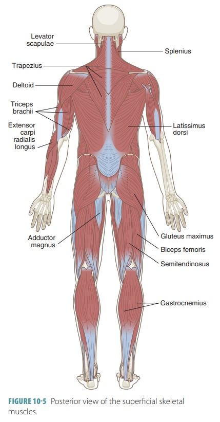
Superficial Skeletal Muscles
There are more than 600 skeletal
muscles in the human body. Superficial muscles are located near the surface of
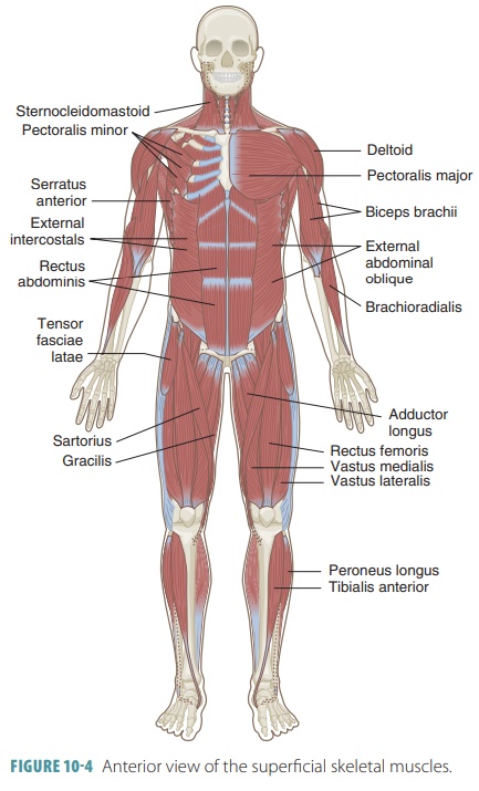
the body. FIGURES 10-4 and 10 -5 show anterior and posterior views of the superficial skeletal muscles.
Axial and Appendicular Muscles
The muscles are subdivided into
the axial and appen-dicular sections. The axial
muscles affect the head, spinal column,
and rib cage. They allow for position-ing of the head and spinal column, move
the rib cage, and assist in the breathing process. They do not move the body or
support the pectoral girdle, pelvic girdle, or limbs. About 60% of the skeletal
muscles are axial muscles. The appendicular muscles stabilize and move the appendicular skeleton, and make up the remaining
40% of skeletal muscles.
The major axial and appendicular muscles are superficial and larger than many other skeletal mus-cles. They cover deeper and smaller muscles that are not visible, unless the overlying muscles are removed or reflected, which means to cut them and pull them out of the way.
It is important to understand the
term innervation, which is the distribution of nerves to a region or organ. For example,
many head and neck muscles are innervated by the cranial nerves. These nerves
begin in the brain, passing through the foramina of the skull. Also, spinal
nerves connect to the spinal cord, passing through the intervertebral foramina.
Spinal nerves may form a complex plexus
or network, after they exit the spinal cord. A single branch of a plexus may
contain axons from several of the spinal nerves.
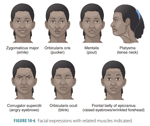
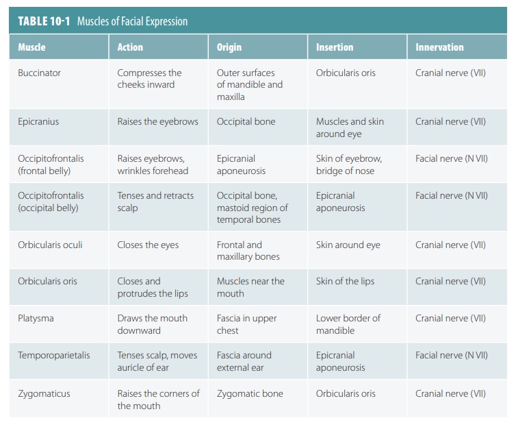
Muscles of Facial Expression
The muscles of facial expression
begin on the sur-face of the skull. At their insertions, the epimy-sium fibers
weave with those of the superficial fascia and the skin’s dermis. The skin
moves when these muscles contract. FIGURE 10-6 shows various facial expressions, listing the muscles that are used to
make them. TABLE 10-1 describes the muscles of facial expression.
Muscles of the Tongue, Pharynx, Larynx, and Palate
The muscles of the tongue have
names that end in glossus. The muscles of the pharynx initiate
the swal-lowing process. The laryngeal muscles control the posi-tion of the
larynx. The muscles of the palate elevate the soft palate and nearby portions
of the pharyngeal wall, and they also pull the entrance to the auditory tube
open. TABLE 10-2 describes the muscles of the tongue, pharynx, and palate.
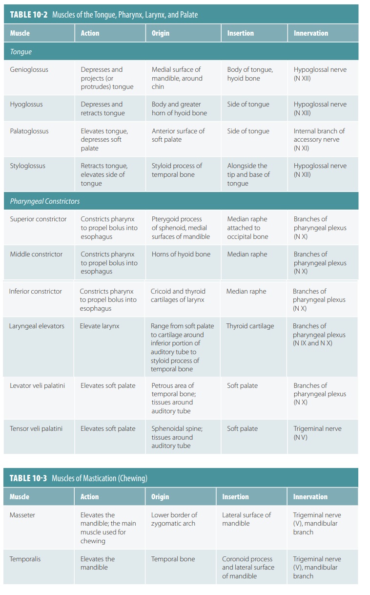
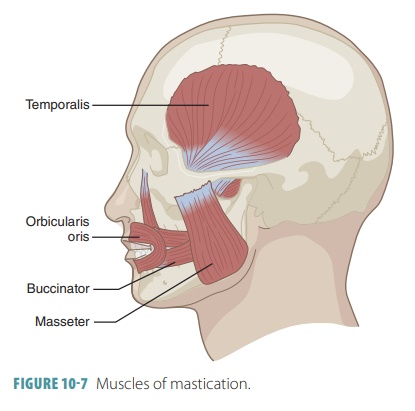
Muscles of Mastication
The muscles of mastication move
the mandible at the temporomandibular joint (TMJ). The strongest jaw muscle is
the masseter. TABLE 10-3 describes the muscles of mastication and FIGURE
10-7 shows these muscles.
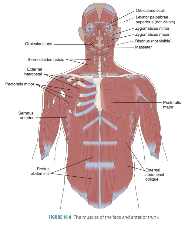
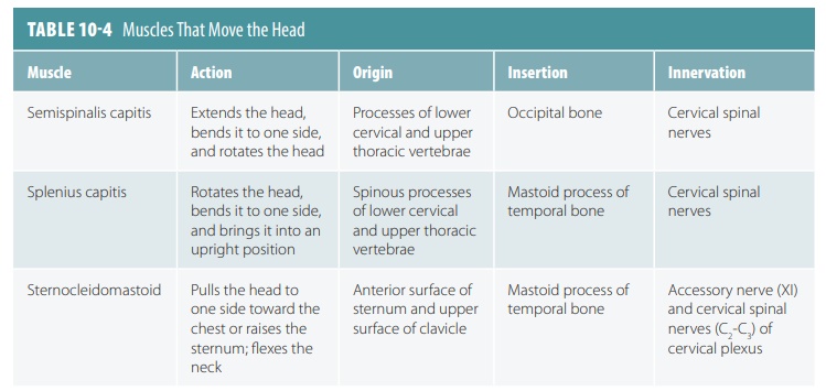
Muscles That Move the Head
The muscles that move the head
originate from the axial skeleton. The major head flexors are the sternocleidomastoid muscles, assisted
by the suprahyoid and infrahyoid muscles. Lat-eral head
movements occur when muscles on just one side of the neck contract. Head
extension is aided by the trapezius
muscles in the back, but the primary extensors of the head are the splenius muscles lying deep below them
(TABLE 10-4). The muscles of the face and anterior trunk are shown in FIGURE 10-8.
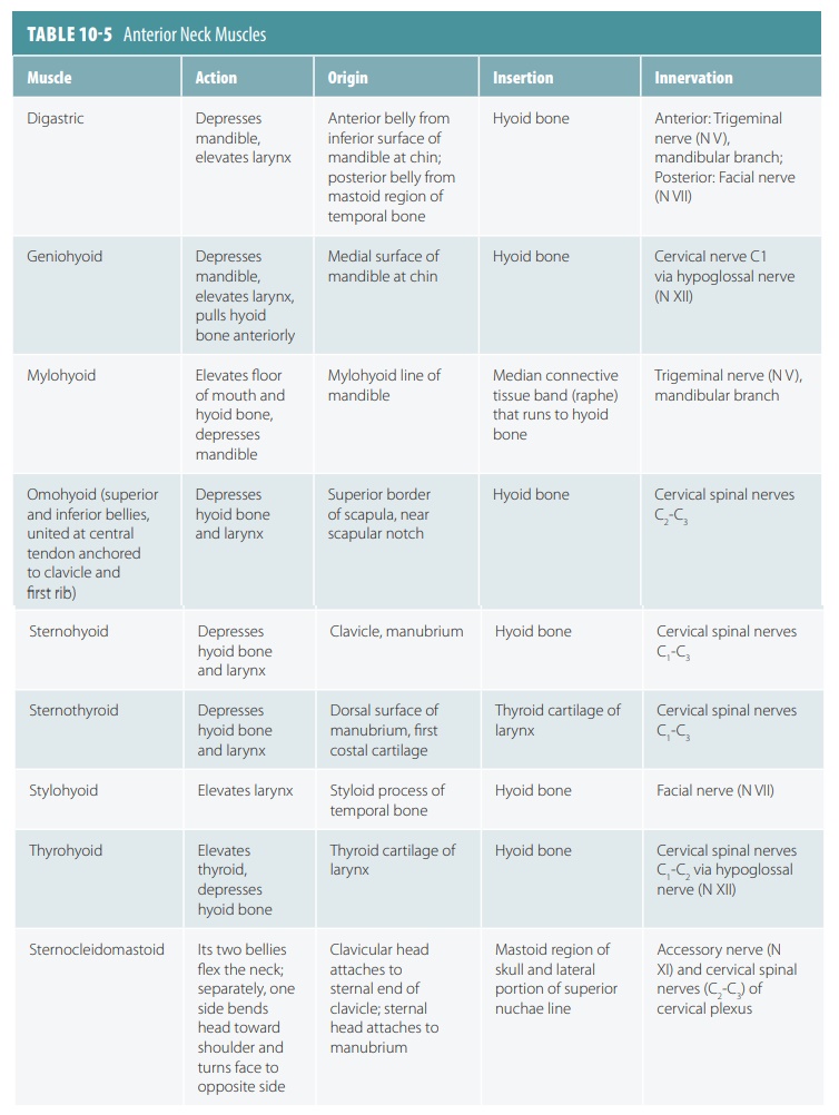
Anterior Neck Muscles
The anterior neck muscles,
described in TABLE 10-5, affect the larynx, mandible, floor of the mouth, tongue, pharynx,
clavicle, sternum, scapula, hyoid, and even the first rib.
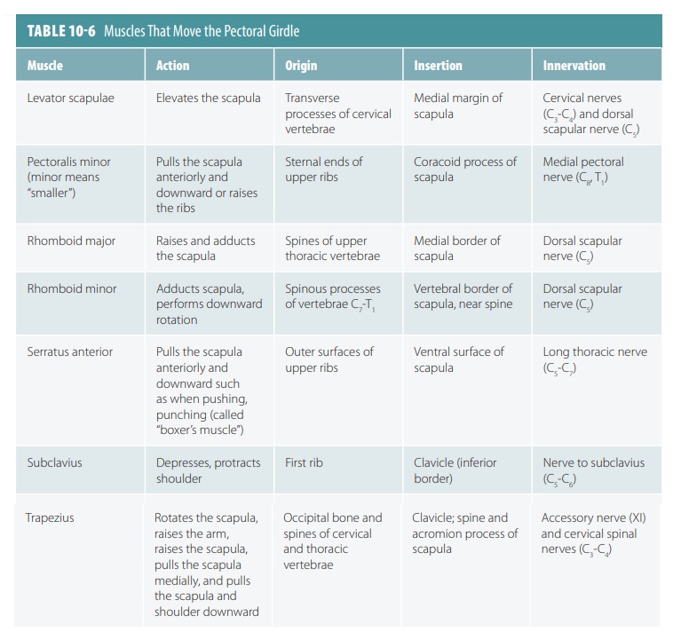
Muscles That Move the Pectoral Girdle
The muscles that move the
pectoral girdle help to elevate, depress, rotate, and move the scapula both laterally
and medially. The clavicles rotate around their axes, providing stability and
precision to the scapula. Most anterior muscles stabilize and depress the
shoulder girdle. The scapular muscle attachments are arranged so that a single
muscle cannot cause a simple linear movement by itself. Instead, several
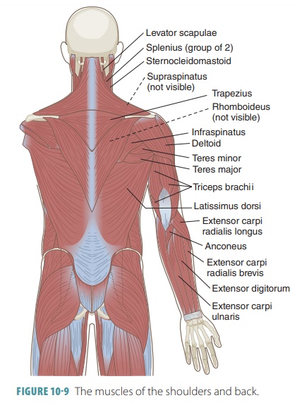
muscles must act together (TABLE 10-6). The muscles of the shoulders and back are shown in FIGURE 10-9.
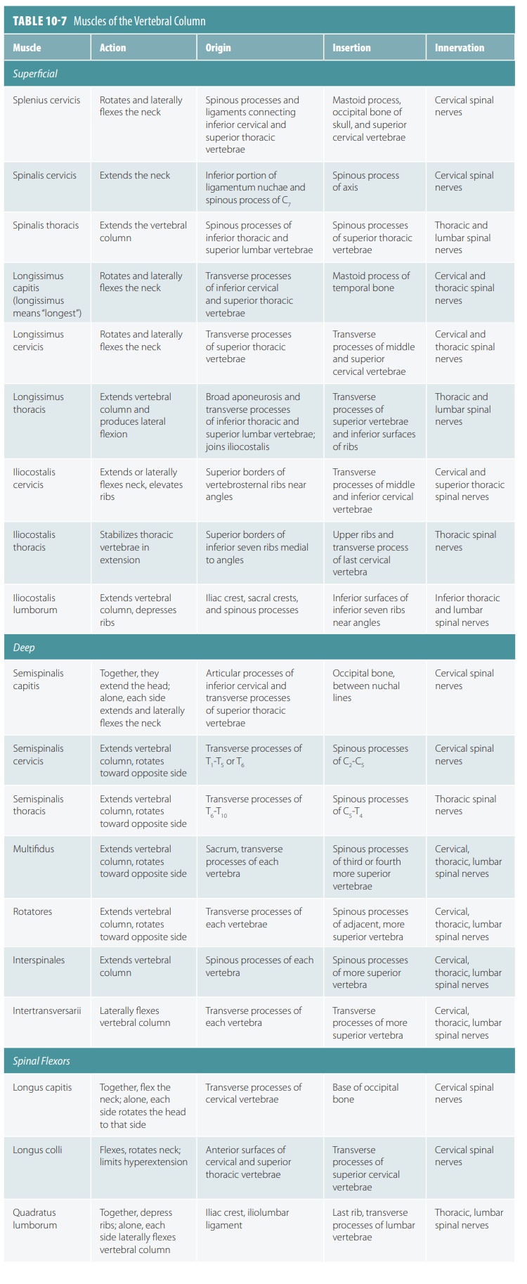
Muscles of the Vertebral Column
The muscles of the vertebral
column lie beneath the more superficial muscles of the back such as the
trapezius and latissimus dorsi (TABLE 10-7). The erec-tor spinae extend the vertebral column when they contract
together. When the muscles on one side con-tract, the vertebral column
experiences lateral flexion.
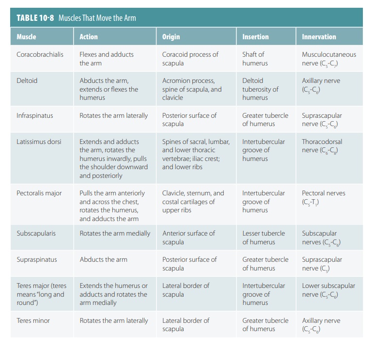
Muscles That Move the Arm
The muscles that move the arm (TABLE 10-8) are
easy to remember since they are grouped by their actions at the shoulder
joints. Of all the arm mus-cles, the deltoid
is the major abductor. The muscles of the anterior shoulders and arms are shown
in FIGURE 10-10.
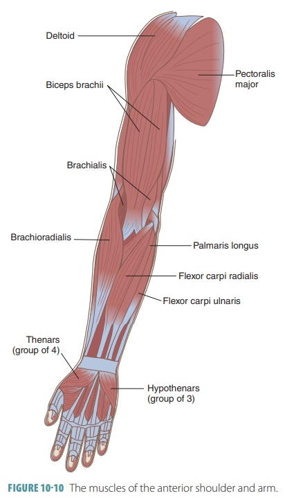
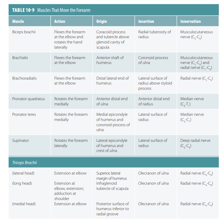
Muscles That Move the Forearm
The muscles that move the forearm
are described in TABLE 10-9 and shown in Figure 10-10. These muscles as well as the
muscles that move the hand mostly orig-inate on the humerus. However, the biceps brachii and the long head of the triceps brachii originate on the
scapula.
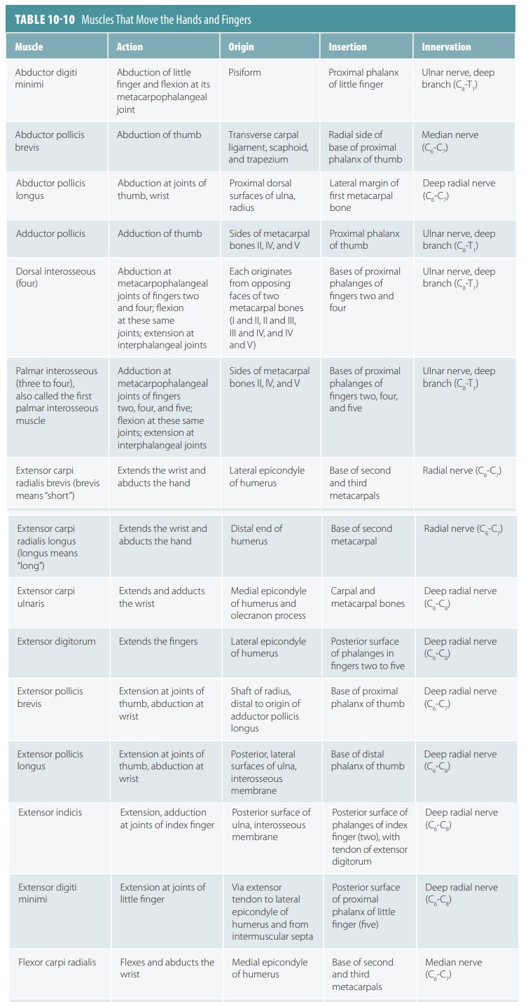
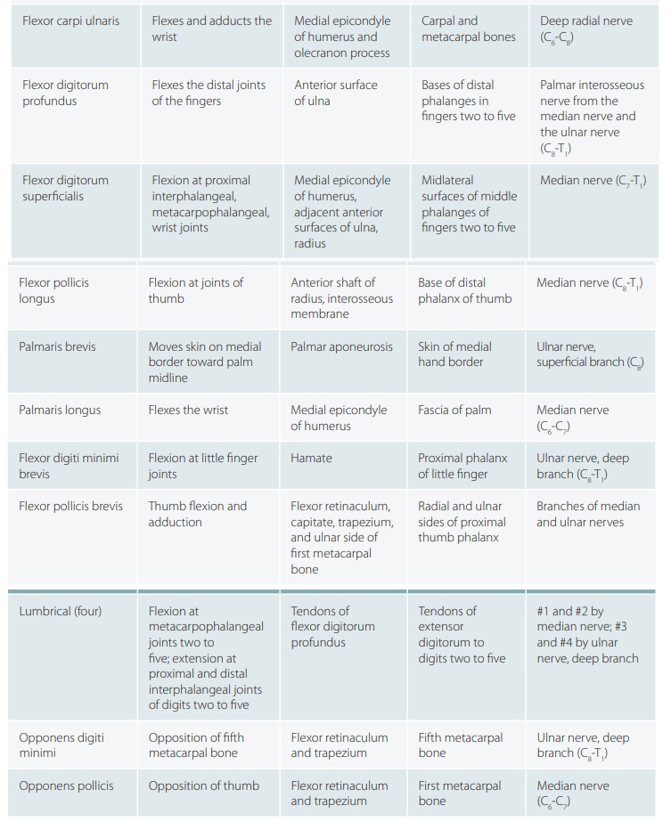
Muscles That Move the Hands and Fingers
Several superficial as well as
deep muscles of the forearm flex and extend the joints of the fingers. They
are large muscles that actually end before reaching the wrists. Their tendons
cross the wrist joints, increasing mobility of the wrists and hands. Strength
and crude control of the hands and fingers are provided by the extrinsic muscles of the forearm,
whereas fine hand control involves the smaller intrinsic muscles originating on the carpal and metacarpal bones.
No muscles originate on the phalanges. The distal joints of the fingers
function by only tendons. The muscles that move the hands and fingers are
dis-cussed in TABLE 10-10.
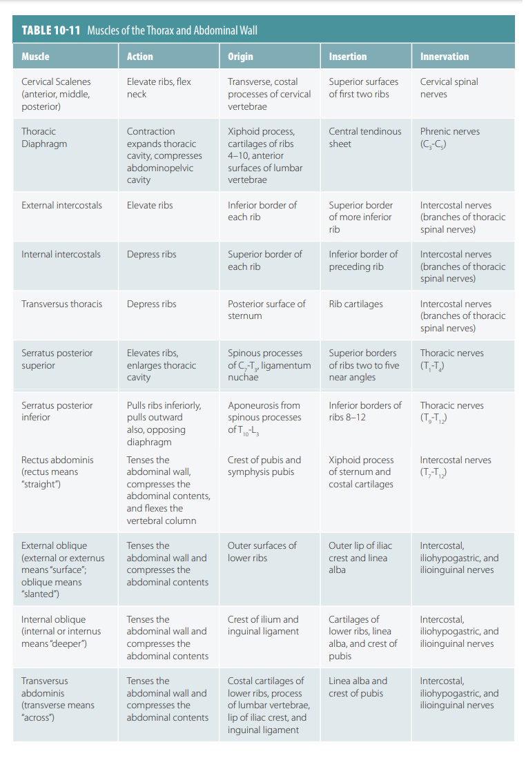
Muscles of the Thorax and Abdominal Wall
The muscles of the thorax and
abdominal wall are described in TABLE 10-11. The deep thorax muscles assist in breathing, with two primary layers
of mus-cles partially forming the anterolateral wall of the thorax. Thoracic
muscles are extremely short and mostly extend from one rib to the next. The
most important muscle of inspiration is the diaphragm.
The anterior and lateral abdominal wall has no ribs, but is composed of four
pair muscles and their related structures. The abdominal wall consists of the
external and internal oblique muscles. The rec-tus abdominis and transversus
abdominis are other major muscles. Together, the abdominal muscles protect and
support the viscera quite well, until they loose muscle tone.
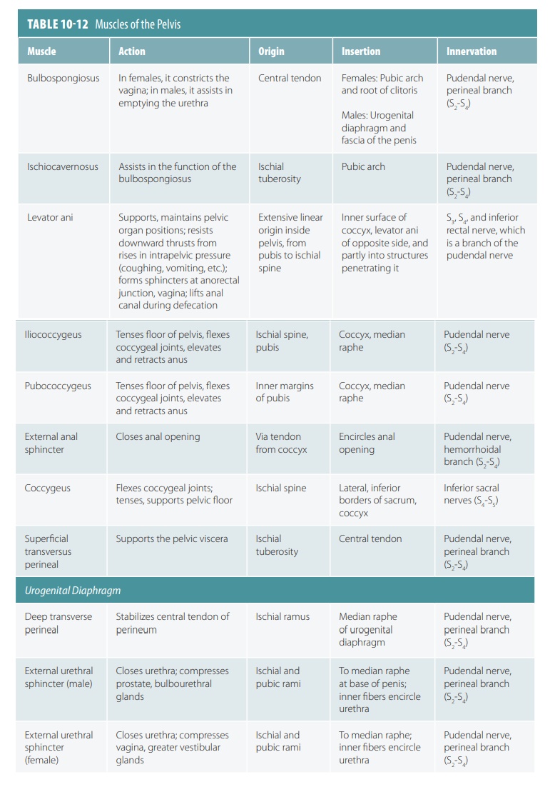
Muscles of the Pelvis
The muscles of the pelvis (TABLE 10-12) are
subdivided into the muscles of the pelvic diaphragm, urogenital diaphragm, and
superficial perineal space.
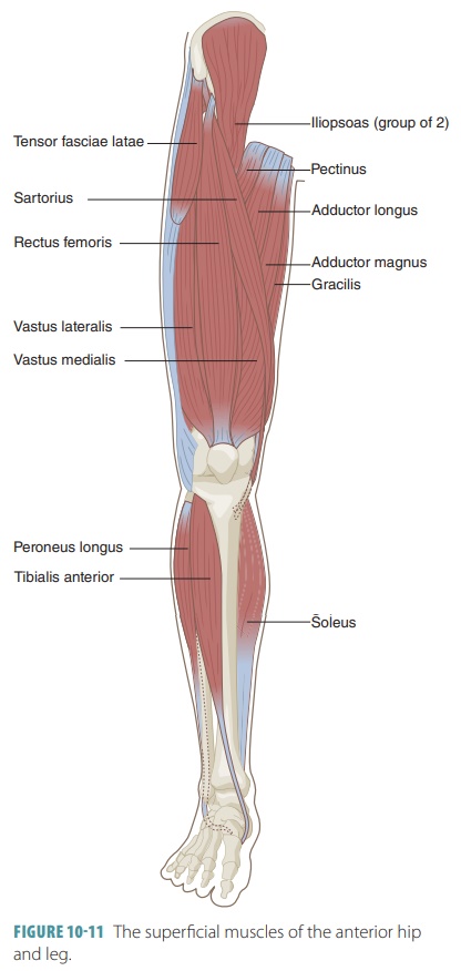
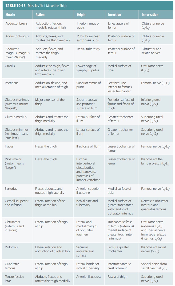
Muscles That Move the Thigh
The muscles of the thigh are
mostly anchored to the pelvic girdle ( TABLE
10-13). The ball-and-socket hip joints
allow flexion, extension, abduction, adduction, circumduction, and rotation.
The related muscles are among the strongest in the body. Most of the thigh
flexors pass in front of the hip joints. The superficial muscles of the
anterior hips and legs are shown in FIGURE 10-11 and the superficial muscles of the posterior hips and legs are shown in
FIGURE 10-12.
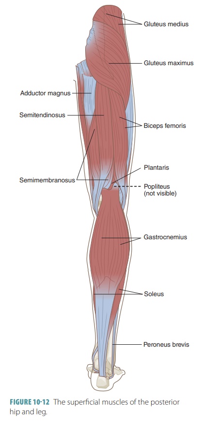
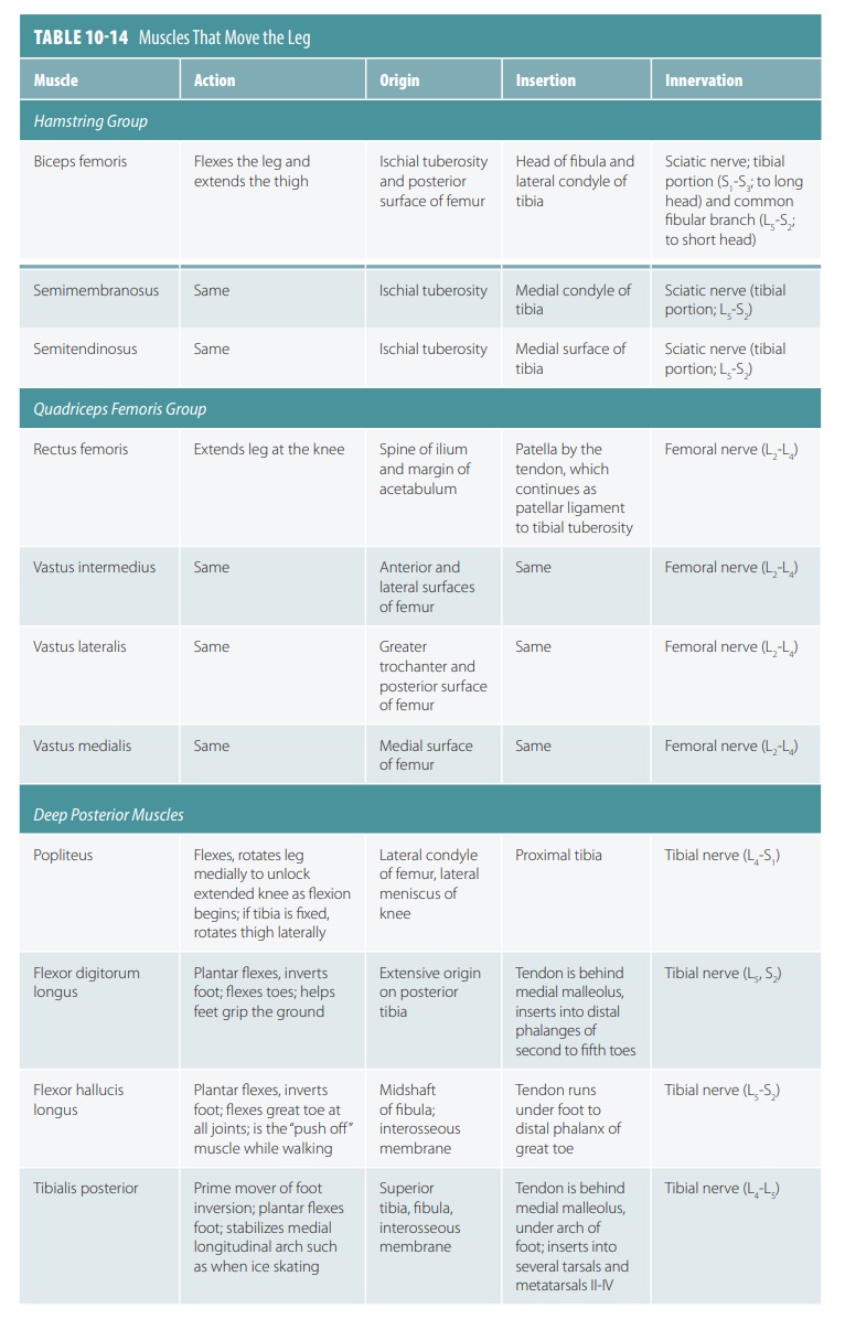
Muscles That
Move the Leg
The muscles that move the leg (TABLE 10-14) allow
for flexion and extension at the knee joint. These muscles may originate on the
pelvis or spine. There are medial, anterior, and posterior compartments of the
thigh. There are also posterior muscles, which include the gluteals and lateral
rotators.
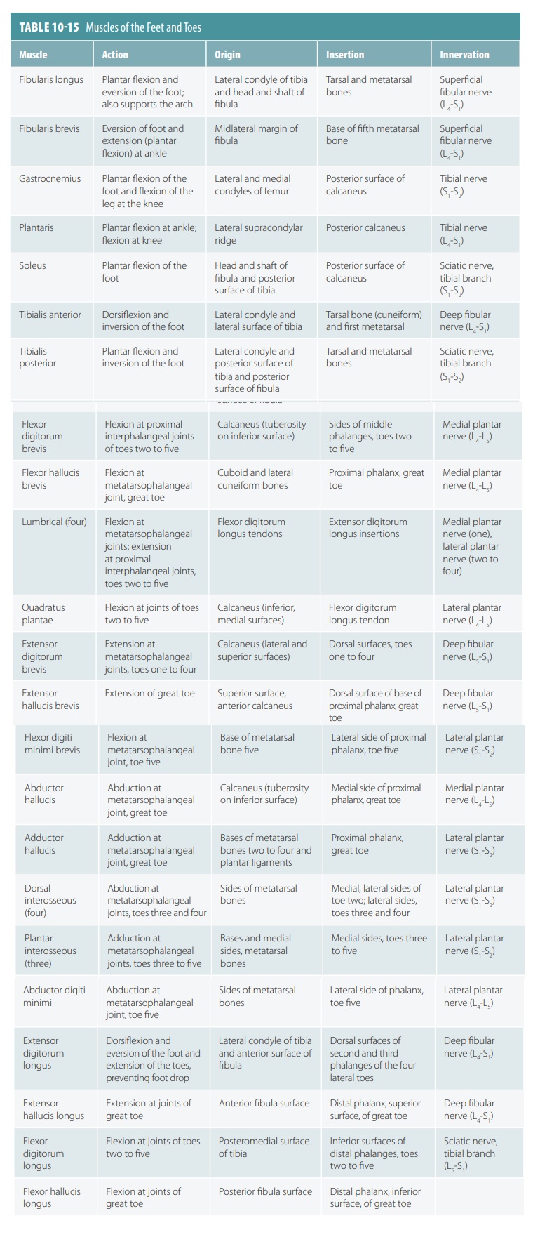
Muscles of the Feet and Toes
The muscles of the feet and toes
are extremely similar to those of the hands and fingers (TABLE 10-15). The
leg muscles that enter the soles. There is a single mus-cle on the foot’s
superior aspect, while there are several muscles on the plantar aspect, in four
layers.
Related Topics