Sarcoplasmic Reticulum and Transverse Tubules
| Home | | Anatomy and Physiology | | Anatomy and Physiology Health Education (APHE) |Chapter: Anatomy and Physiology for Health Professionals: Support and Movement: Muscle Tissue
Inside the sarcoplasm of a muscle fiber, a network of channels surrounds each myofibril.
Sarcoplasmic
Reticulum and Transverse Tubules
Inside the sarcoplasm of a muscle
fiber, a network of channels surrounds each myofibril. These membranous
channels form the SR, which has interconnected tubules surrounding each
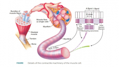
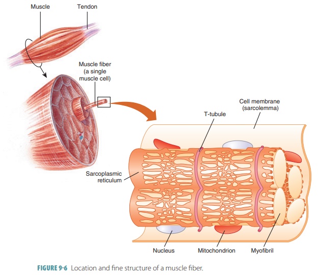
myofibril (FIGURE 9-6). Most of these tubules run longitudinally and communicate at the H
zone.
Sarcoplasmic Reticulum
Each skeletal muscle fiber
contains transverse tubules as well as a SR,
which is a complex smooth endoplas-mic reticulum. It has interconnecting
tubules that surround each myofibril. Most of these tubules run lengthwise
along the myofibrils and communicate with them at the H zone. However, the terminal cisterns form large cross-channels that are perpen-dicular, located at the
A band- I band junctions. These always occur in pairs. Large numbers of
glycogen granules and mitochondria are involved in produc-ing energy needed for
muscle contractions. The SR regulates intracellular ionic calcium levels. It
stores calcium and also releases it as needed, when muscle fibers are
stimulated to contract. Calcium provides the final “start” signal for muscle
contraction. Inte-gral proteins protrude into the intermembrane spaces from the
SR and T-tubules. These proteins act to sense
voltages. The SR proteins form gated channels through which calcium ions are
released by the ter-minal cisterns.
Transverse Tubules
Transverse
tubules (T-tubules) are other
mem-branous channels extending inward and passing through the fiber. Action
potentials are conducted into a skeletal muscle fiber by T-tubules. These
tubules open to the outside of the muscle fiber and contain extracellular
fluid. Each tubule lies between enlarged structures called terminal cisternae,
near the point where actin and myosin filaments over-lap. Together, the SR and
T-tubules activate muscle contraction when stimulated. The functional role of
the T-tubules is to enhance cellular communica-tion during muscle contraction.
The lumen of each T-tubule is continuous with the extracellular space. The T-tubules form triads , which are
groupings consisting of themselves in between two terminal cisterns. Ultimate
control of muscle contraction is via electrical impulses initiated by nerves
that travel through the sarcolemma. The continuous T-tubules conduct impulses deeply into muscle cells and all sarcomeres. This
signals the release of calcium from nearby terminal cisterns. At the triads,
organelles are in close contact and integral proteins protrude into
intermembrane spaces to act as voltage sensors. The proteins in this location
that derive from the SR form gated channels. Through these channels, ter-minal
cisterns release calcium ions.
1. Describe
action potential generation and propagation.
2. Identify
the functions of the transverse tubules and SR.