Neuromuscular Junction
| Home | | Anatomy and Physiology | | Anatomy and Physiology Health Education (APHE) |Chapter: Anatomy and Physiology for Health Professionals: Support and Movement: Muscle Tissue
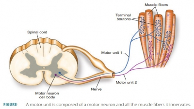
Somatic motor neurons are nerve cells that activate skeletal muscle fibers.
Neuromuscular
Junction
Somatic motor neurons are nerve
cells that activate skeletal muscle fibers. They are found in the brain and
spinal cord, but have long, thread-like extensions (axons) that are connected
inside nerves to muscle cells they serve. Each axon ending forms an elliptical neuromuscular junction (end plate) with just one muscle fiber. Axon
terminals and muscle fibers are
separated by a space called the synaptic
cleft. Inside each axon terminal
are synaptic vesicles, which are membranous sacs that
contain ACh. Junctional folds of each
sarcolemma pro-vide large surface areas for the millions of nearby ACh
receptors. Therefore, neuromuscular junctions include axon terminals, synaptic
clefts, and junctional folds.
To understand more completely,
the steps in which a motor neuron stimulates a skeletal muscle fiber, follow:
■■ A nerve impulse reaches the end of an axon, and the axon terminal
releases ACh into the synaptic cleft. Calcium ions activate synaptic vesicles.
■■ ACh diffuses across the cleft, attaching to ACh receptors on the muscle
fiber’s sarcolemma.
■■ Binding of ACh triggers electrical events that gen-erate an action
potential.
The effects of ACh are quickly
terminated by acetylcholinesterase,
which breaks down ACh into its basic
elements (acetic acid and choline). Therefore, continued and undesirable muscle
fiber contraction, without additional nervous system stimulation, is prevented.
Related Topics