Joint Injuries
| Home | | Anatomy and Physiology | | Anatomy and Physiology Health Education (APHE) |Chapter: Anatomy and Physiology for Health Professionals: Support and Movement: Articulations
Sprains and dislocations are the most common joint injuries, but cartilage tears are commonly seen joint injuries in athletes.
Joint
Injuries
Sprains and dislocations are the
most common joint injuries, but cartilage tears are commonly seen joint
injuries in athletes. In a sprain, there is stretching or tearing of the ligaments that reinforce a
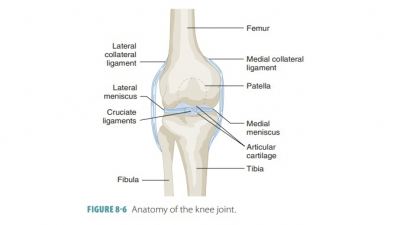
joint. The most commonly sprained joint ligaments are those of the ankle, knee,
and lumbar spine. Sprains are usually painful and cause the immobilization of
the injured patient. When partially torn, they heal very slowly because of a
lack of vascularization. However, a com-plete ligament tear is treated with
surgery, grafting, and long-term immobilization. Ends of ligaments can be
sutured together, but this is difficult to perform because of the hundreds of
fibrous strands involved in each ligament. Grafting is used instead for ligaments
such as the anterior cruciate ligament. In this oper-ation, part of a muscle
tendon is attached to articu-lating bones. For other ligaments (such as the
medial collateral ligament of the knee), long-term immobili-zation is as
effective as surgical methods.
A dislocation is also known as a luxation.
It occurs when bones are forced out of alignment and usually is involved with a
sprain. There is inflam-mation and difficulty in moving the joint. Common
causes of dislocations include falling and contact sports. The most commonly
dislocated joints are those of the jaw, fingers, thumbs, and shoulders.
Dislocations, like fractures, must be reduced.
This means the ends of the bones must be returned to their proper positions by
a physician. Partial dislo-cation of a joint is called subluxation. Because an ini-tial dislocation stretches a joint’s
capsule as well as its ligaments, repeat dislocations of the same joint often
occur. The joint then has poor reinforcement because the capsule has become
loose.
Tearing of cartilage causes it to
break or pop because of being overstressed. The most common areas of torn
cartilage occur in the knee menisci. Usually, the meniscus receives a
compression and shear stress simultaneously, resulting in tearing. Cartilage
usually remains torn because it cannot usually and sufficiently repair itself.
Loose bodies (fragments of cartilage) interfere with joint function because
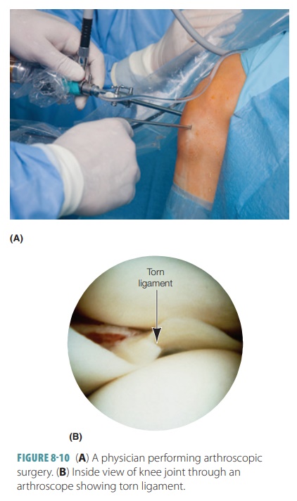
they cause bind-ing or locking of the joint. Therefore, damaged carti-lage is
usually surgically removed via arthroscopic surgery (FIGURE 8- 10). Fortunately, the patient is
usually able to leave the hospital the
same day as the surgery. An arthroscope
is used, which is very small, containing a miniscule lens and fiberoptic light
source. The surgeon can look inside the joint to determine sur-gical options.
Ligaments can be repaired or fragments of cartilage removed through one or
several small slits. This reduces tissue damage and scarring. If only part of
the meniscus is removed, mobility is not severely impaired, but the joint
becomes much less stable. If the entire meniscus is removed, osteoarthritis usu-ally develops in the joint earlier than normal. A menis-cal
transplant may be used for younger patients when cartilage is extensively
damaged. Future surgeries may involve transplantation of a patient’s own stem
cells.
Related Topics