Platelets or thrombocytes
| Home | | Anatomy and Physiology | | Anatomy and Physiology Health Education (APHE) |Chapter: Anatomy and Physiology for Health Professionals: Blood
Platelets in nonmammalian vertebrates are nucle-ated cells called thrombocytes.
Formed Elements
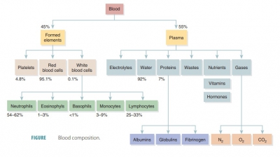
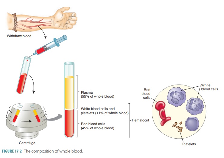
The formed elements of blood include erythrocytes, leukocytes, and platelets. Red blood cells make up about 45% of blood volume, which is known as the hematocrit. The RBCs have no nuclei or organelles,meaning that they are not considered “true cells.” Platelets are only cell fragments. Only leukocytes are complete cells. Leukocytes and platelets make up less than 1%. The remainder is plasma. Most blood cells do not divide. Instead they are replaced when stem cells continuously divide in the bone marrow. All formed elements arise from the hematocytoblasts, also called hematopoietic stem cells, which are undifferen-tiated precursor cells in the red bone marrow. Most formed elements only survive in the bloodstream for a few days. Different modes of maturation of formed elements exist. Once a cell becomes committed to a certain blood cell pathway, it is unable to change. Membrane surface receptors appear, which signal the cell’s commitment to one blood cell pathway. The receptors respond to specific growth factors or hor-mones. These assist the cell in becoming even more specialized. In a healthy male adult the normal hema-tocrit value is 47%, plus or minus 5%. In a healthy female adult, it is 42%, plus or minus 5%. Less than 1% of blood volume consists of platelets and leukocytes. Most of the remaining 55% of whole blood is made up by the plasma (FIGURE 17- 2). The components that make up whole blood can be separated or fractionated to be clinically analyzed.
Platelets
Platelets
in nonmammalian vertebrates are nucle-ated cells called thrombocytes. They are incomplete
cells arising from extremely large red bone marrow cells called megakaryocytes that have become
frag-mented. Megakaryocytes, which are up to 60 μm in diameter, develop
from hemocytoblasts because of the hormone thrombopoietin. Plasma membranes from
each megakaryocyte fragment seal quickly around their cytoplasm to form a
platelet. The formation of platelets is known as thrombocytopoiesis. Platelets lack nuclei and are approximately
one-fourth of the size of a lymphocyte and less than one-half the size of an
erythrocyte. In actual size, platelets measure between 2 and 4 μm
in diameter. They live for about 10 days and are capable of amoeboid motion.
Usually, platelet counts range from 150,000 to 400,000 per μL.
They can circulate freely and are inactivated during this activity by
prostacyclin and nitric oxide from endo-thelial cells of the blood vessels.
Approximately one-third of the body’s platelets are held in the spleen and
other vascular organs instead of in the bloodstream. These reserves are
mobilized during a circulatory cri-sis such as severe bleeding. The function of
platelets is primarily to block injuries to damaged blood vessels and to start
forming blood clots. They accomplish this by sticking to the damaged site and
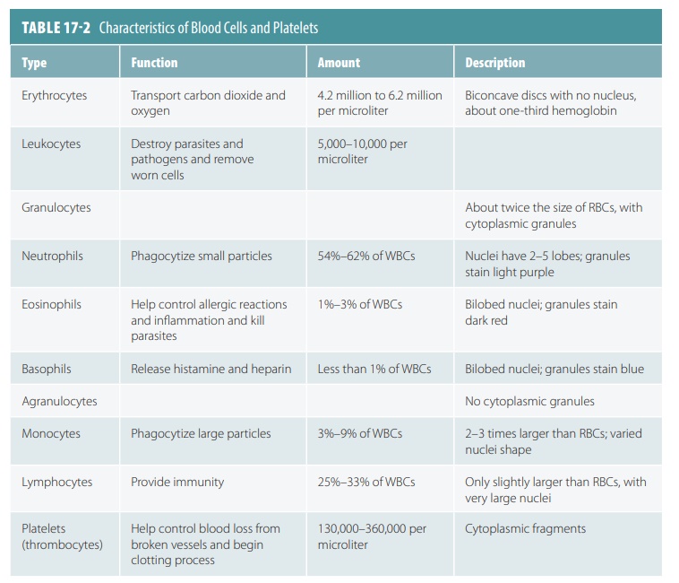
forming a tempo-rary plug to seal the broken area. The characteristics of RBCs,
WBCs, and platelets are listed in TABLE
17-2.
When a blood smear is performed, each platelet’s outer region stains blue, whereas the inner area has granules that stain purple. The granules contain many chemicals used for clotting, including enzymes, adenosine diphosphate, platelet-derived growth factor, calcium ions, and serotonin.
1. Name the three major types of blood cells.
2. Explain how platelets participate in the clotting process.
3. Describe thrombopoietin.
4. What is a
megakaryocyte?