Synovial Joints
| Home | | Anatomy and Physiology | | Anatomy and Physiology Health Education (APHE) |Chapter: Anatomy and Physiology for Health Professionals: Support and Movement: Articulations
These joints allow free movement and are referred to as diarthrotic. They are more complex than other types of joints.
Synovial Joints
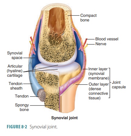
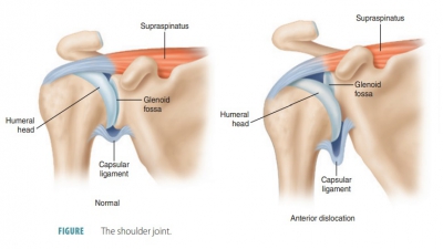
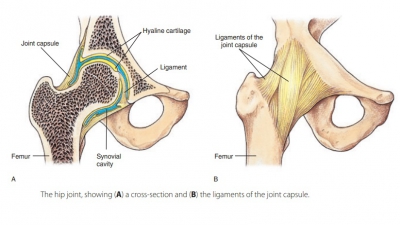
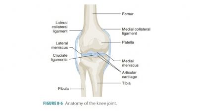
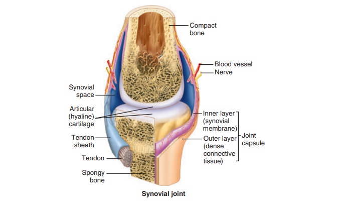
These joints allow free movement and are referred to as diarthrotic. They are more complex than other types of joints. Synovial joints have outer layers of ligaments (joint capsules) and inner linings of synovial membrane that secrete synovial fluid, which lubricates these joints (FIGURE 8-2). Some synovial joints have shock-absorbing fibrocartilage pads called menisci. They may also have fluid-filled sacs (bursae), commonly located between tendons and underly-ing bony prominences such as in the knee or elbow. The synovial fluid lubricating these joints allows for great freedom of movement. All synovial joints are freely moving diarthroses. Most joints of the body and nearly all joints of the limbs are synovial joints. Fat pads are localized adipose tissue masses that are covered by a synovial membrane layer. The six character-istics of synovial joints are:
■■ Articular cartilage: This is made up of smooth hyaline cartilage covering opposing bone surfaces. Although only one millimeter or thinner across, articular cartilage absorbs compression and helps to keep bone ends from being crushed.
■■ Articular cavity: This is a potential space containing a small amount of synovial fluid.
■■ Articular capsule: This two-layered structure that encloses the joint cavity has a tough outer fibrous layer made up of dense irregular connective tissue, which is continuous with the periostea of articulating bones. The capsule strengthens the joint and ensures the bones are not pulled apart. Each joint capsule’s inner layer is known as the synovial membrane, made of loose connective tissue that lines the fibrous layer’s internal portion and covers all internal surfaces of joints that are not covered with hyaline cartilage. The synovial membrane manufactures the synovial fluid.
■■ Synovial fluid: This slippery liquid found in all free spaces inside the joint capsule is mostly syn-thesized by filtration from blood in the synovial also found inside articular cartilages as a slippery film. It bears weight, reducing friction between cartilages. Without it, joint surfaces would be destroyed from use. When a joint is compressed, the fluid is forced out of the cartilages. As joint pressure is relieved, it flows back into the articular cartilages quickly. This process is called weeping lubrication, which also provides nourishment to the cells of the joint cartilage. Synovial fluid also has phagocytic cells that patrol the joint cavity for microbes and cell debris.
■■ Reinforcing ligaments: These are band-like acces-sory structures that reinforce and strengthen syno-vial joints. They are primarily capsular ligaments (actually, thicker parts of the fibrous layer). They are distinct from and remain outside the capsule (extracapsular ligaments) or remain deep to it (intracapsular ligaments). The intracapsular lig-aments do not actually lie within the joint cavity because they are covered with synovial membrane. The term double-jointed actually means a person’s joint capsules and ligaments are looser, with more ability to stretch, than those of the average person.
■■ Nerves and blood vessels: Plentiful in synovial joints, sensory nerve fibers innervate the joint cap-sule, whereas most of the blood vessels supply the synovial membrane. Some sensory nerve fibers detect pain, but most regulate the joint’s position and stretching ability. Therefore, these fibers allow the nervous system to monitor body posture and movements. Extensive capillary beds produce the blood filtrate, which is the basis of synovial fluid Synovial joints sometimes have other structural components. For example, the knee and hip joints have fatty pads between the fibrous layer and the synovial membrane or bone. These add extra cushioning. Other synovial joints have fibrocartilage wedges or discs that separate articular surfaces (these menisci or articular discs were introduced earlier). They extend in from the articular capsule, dividing (or partially dividing) the synovial cavity in half. Articular discs help articu-lating bone ends to fit better, stabilizing the joint and lessening friction on the joint surfaces. Articular discs occur in the knees, jaw, and other areas.
