Fibrous Joints
| Home | | Anatomy and Physiology | | Anatomy and Physiology Health Education (APHE) |Chapter: Anatomy and Physiology for Health Professionals: Support and Movement: Articulations
Describe fibrous joints and list three examples.
Fibrous
Joints
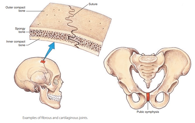
Lying between bones that are in
close contact with each other, fibrous
joints are joined by a thin, dense
connective tissue (FIGURE 8-1). They have no actual joint cavity. An example of a fibrous joint is a
suture between flat bones of the skull. No real movement takes place in most
fibrous joints, making them synar-throtic in classification. Those with limited
movement (amphiarthrotic) include the joint between the distal tibia and
fibula. The amount of movement they have depends on the length of the
connective tissue fibers that unite the bones. There are three types of fibrous
joints: sutures, syndesmoses, and gomphoses.
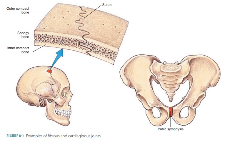
Sutures
Sutures are seams that occur only between the bones of the skull. They
have waved and articulated bone edges that interlock. Each junction is totally
filled by a tiny amount of extremely short connective tissue fibers. These
fibers are continuous with the periosteum, creat-ing rigid structures joining
the bones together. However, they also allow the skull to expand during childhood,
when the brain is growing. Closed sutures, during brain growth, are better
described as synostoses. The immo-bility of the sutures helps to protect the brain.
Syndesmoses
In syndesmoses, ligaments connect the bones and the connecting fibers are longer than
those found in sutures. The varied lengths of these fibers control the amount
of movement that can occur. Syndesmoses with shorter fibers have little or no
allowed “give” (movement), for example, the ligament connecting the distal ends
of the fibula and tibia. When they are longer, more movement is possible, for
example, the interosseous membrane (similar to a ligament) that connects the
ulna and radius.
Gomphoses
Gomphoses are fibrous joints with a peg-in-socket structure. In the
human body, gomphoses are only exemplified by the articulation of the teeth in
their alveolar sockets. The singular term gomphosis
refers to how the teeth are embedded in their socket (as if they were hammered
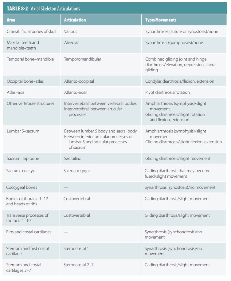
in) . In gomphoses, the fibrous connects are the short periodontal ligaments. Articulations of the axial skeleton are described in TABLE 8-2.
1.
Describe fibrous joints and list three examples.