Pelvic Girdle - Appendicular Skeleton
| Home | | Anatomy and Physiology | | Anatomy and Physiology Health Education (APHE) |Chapter: Anatomy and Physiology for Health Professionals: Support and Movement: Bone Tissues and the Skeletal System
1. Which bones are involved in the formation of the hip bone? 2. List the bones of the lower limbs. 3. Which bone is the strongest in the human body? 4. Which arches of the feet support the weight of the body?
Appendicular
Skeleton
The appendicular skeleton
contains the upper and lower limb bones and the bones anchoring the limbs to
the axial skeleton. The appendicular skeleton includes the pectoral girdle,
upper limbs, pelvic girdle, and lower limbs.
Pelvic Girdle
Two hip
bones, which articulate with each
other and the sacrum, make up the pelvic girdle (FIGURE 7-32). The hip bones are also called the coxal or pelvic bones. The pelvic
girdle attaches the lower limbs to the axial skeleton. Together, the sacrum,
coccyx, and pelvic girdle form the pelvis.
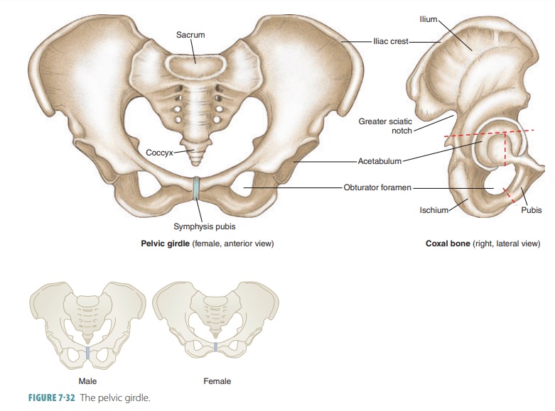
The pelvis of females is usually
wider in all diam-eters than that of males. The pelvic girdle supports,
protects, and/or articulates with the trunk, lower limbs, urinary bladder,
large intestine, and repro-ductive organs. The hip bones each have three parts
(the ilium, ischium, and pubis) fused together into an acetabulum, which houses the rounded head of the
femur (thigh bone).
Ilium
The largest portion of the hip
bone is the ilium , form-ing the prominence of the hip. The margin of the prominence is
called the iliac crest. This crest ends anteriorly in a blunt anterior superior iliac spine and ends posteriorly in a sharp posterior superior iliac spine. The anterior superior iliac spine
is an import-ant landmark since it is easily felt through the skin, and is even
visible when an individual is thin. Just inferior to the posterior inferior
iliac spine there is a deep indentation in the ilium, which forms the greater sciatic notch. Through this notch, the thick
and rope-like sciatic nerve passes to
enter the thigh. The posterolateral surface of the ilium is broad and is called
the gluteal surface. Three ridges cross this surface, which are known as the posterior, anterior, and inferior gluteal lines. The gluteal (buttock)
mus-cles attach to these lines.
The ilium is made up of a body and a superior, wing- shaped
portion called the ala. The body of
the ilium joins the pubis anteriorly and joins the ischium inferiorly. The
ilium joins the sacrum at the sacroiliac joint. A projection from the ilium
provides attach-ments for ligaments and muscles. Ligaments from the iliac tuberosity
stabilize the sacroiliac joint. Each sacroiliac joint allows the body’s weight
to be transmitted from the spine to the pelvis. A promi-nent arcuate line runs
inferiorly and anteriorly from the auricular surface. It is a ridge that helps
cre-ate the pelvic brim, which is the
superior margin of the true pelvis.
Ischium
The ischium is the lowest portion of the hip bone and is L-shaped. Its angle, the
ischial tuberosity, points downward and posteriorly. Each ischial tuberosity is
very strong and thick. It supports the body weight when sitting. There are two
other important areas of the ischim: the ischial
spine and the lesser
sciatic notch
. The ischial spine projects medially
into the pelvic cavity. It is a point of attachment of the sacrospi-nous ligament that runs from
the sacrum. Just inferior to this is
the lesser sciatic notch, through which many nerves and blood vessels pass to
supply the anogenital region. The inferior surface of the ischial body forms
the ischial tuberosity, having a rough texture.
Pubis
The pubis is the anterior portion of the hip bone, and forms an angle known as
the pubic arch or subpubic angle. This arch helps to differentiate between the pelvises of males and females. The
pubis is basically V-shaped, with superior and inferior rami extending from its flattened, medial body. The pubic crest is formed by the thickened anterior border. The lateral end of the
pubic crest is the pubic
tubercle, which is one of the attachments
for the inguinal ligament. The two
pubic bones join at the symphysis pubis,
the upper margin of which (the pelvic brim) separates the lower pelvis from the
upper portion. A large open-ing, known as the obturator
foramen, lies between the pubis and
ischium. It is nearly closed by a fibrous membrane.
Lower Limbs
The lower
limbs consist of the bones of the
thigh, leg, and foot. In each lower limb, these bones include a femur, a
patella, a tibia, a fibula, tarsals, metatarsals, and phalanges.
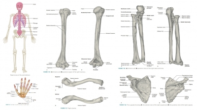
Femur
The femur or thighbone is the longest, strongest, and largest bone in the body,
extending from the hip to the knee (FIGURE 7-33). Various processes from the femur provide attachments for muscles of
the lower limbs and buttocks. A pit on the head of the femur called the fovea capitis marks
the point of attach-ment of the ligamentum capitis. Just below the head is a
neck (constriction) and large processes known as the lateral greater trochanter and the
medial lesser trochanter, where the thigh and buttock
muscles are attached. The trochanters
are connected anteriorly by the intertrochanteric
line and connected posteriorly by the prominent intertrochanteric crest.
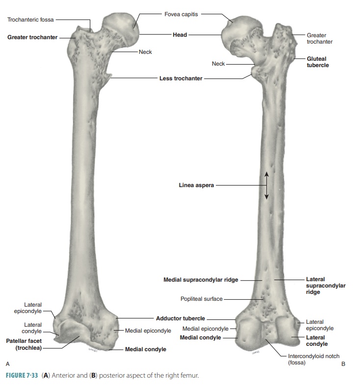
Proximally, the femur articulates with the hip bone, coursing medially over its length toward the knee. Because of this structure, the knee joints can remain closer to the body’s center of gravity, providing better balance. In females, the medical course of each femur is more pronounced, due to their pelvises being wider. This can cause a higher likelihood of knee problems in females who are athletic. The femur, after broadening distally, articulates with the tibia via the lateral and medial condyles, which are processes flanked superiorly by lateral and medial epicondyles or sites of muscle attachment. The superior region of the medial epicondyle has a bump known as the adductor tubercle. Between the condyles on the anterior femoral surface, the smooth patellar surface articulates with the patella. There is a deep, U-shaped intercondylar fossa between the condyles on the posterior aspect of the femur.
Patella
The kneecap (patella) is a
triangular-shaped bone that articulates with the femur and is located in a
ten-don passing over the knee. The patella has a convex, rough anterior surface
and a broad base. The apex of the patella is connected to the tibia by the
patellar lig-ament. The rough surface of the patella is where the anterior and
superior surfaces of the quadriceps ten-don and the patellar ligament attach.
Tibia
The tibia or shinbone, is the larger of
the two leg bones, located on the medial side. Its distal end expands to form a
prominence on the inner ankle where liga-ments attach (FIGURE 7-34). A
depression on its lateral side articulates with the fibula. The tibial tuberosity is the
process where the patellar ligament attaches. The distal end of the tibia has
an inner prominence (the medial malleolus)
where ligaments attach.
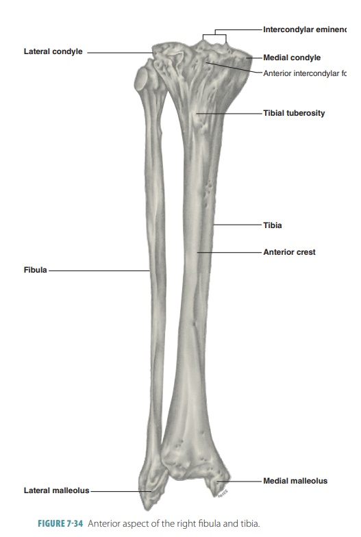
Fibula
The fibula is a slender bone located on the lateral side of the tibia. It does not
enter into the knee joint and does not bear any body weight. The head of the
fibula articulates with the tibia. The fibula has slightly enlarged ends, a proximal
head and a distal lateral
malleolus.
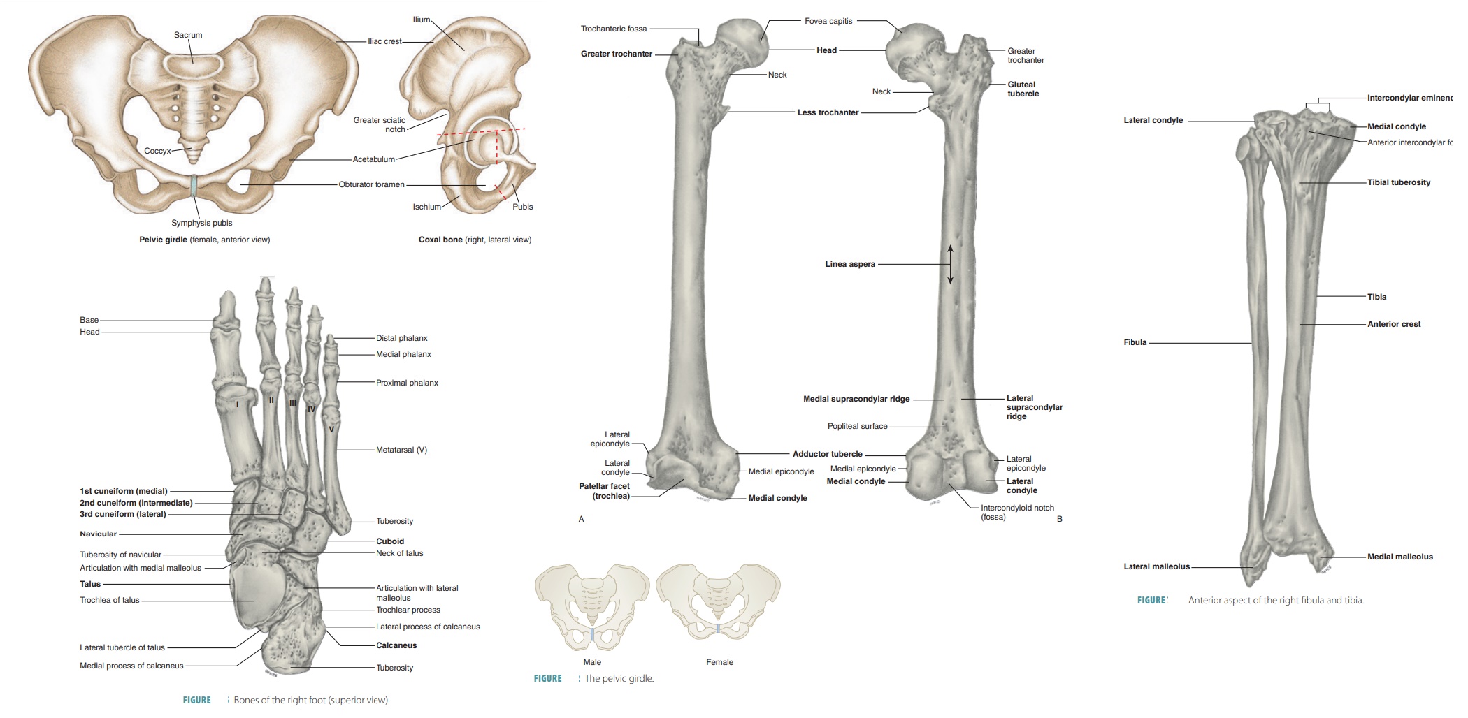
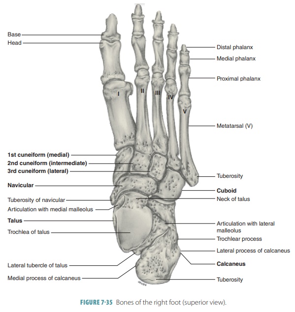
Foot
The foot includes the ankle, instep, and toes. The ankle or tarsus is made up of seven bones called
tarsals that are arranged so the
talus bone moves freely where it joins
the leg bones. The tarsal bones connect the tibia and fibula to the foot. The
other tarsal bones are firmly bound in a mass supporting the talus. The largest
tarsal bone is the calcaneus(heel bone), which helps support
body weight and provides muscle attachment for foot movement.
The Achilles tendon attaches to
the calcaneus. The other tarsals are the lateral cuboid, the medial navicular, and the anterior medial, intermediate, and lateral cuneiform bones.
The cuboid and cuneiform bones articulate with the metatarsal bones
anteriorly.
Metatarsals
The instep (metatarsus) is made
up of five bones called metatarsals numbered one through five, beginning with the medial side.
These bones are small and long, except for the first metatarsal, which is thick
and short, playing an important role in supporting the
weight of the body. The metatarsals have a more par-allel arrangement than the
metacarpals in the hands. The “ball” of the foot is formed by the enlarged head
of the first metatarsal. It articulates distally with the proximal phalanges of
the toes.
Phalanges
The 14 phalanges of the toes,
similar to those of the fingers, are aligned with the metatarsals. Each toe has
three phalanges except the great toe or hallux,
which has only two—proximal and distal (FIGURE 7 -35). The
phalanges of the toes are smaller than the phalanges of the fingers and have
less mobility, though they are similar in structure and arrangement.
Arches of the Foot
The two longitudinal arches of the foot allow for weight transfer to occur
and are maintained by the ligaments and tendons that bind the calcaneus to the
distal portions of the metatarsal bones. The degree of curvature from the
medial to lateral foot borders is called the transverse arch. The foot bones interlock , which along with the
strength of the ligaments and the pull of certain tendons maintains the arches
during muscular activity. The arches stretch as needed when walking or running.
The medial longitudinal arch curves significantly because of the position of the talus. The lateral longitudinal arch is lower, elevat-ing the lateral foot to redistribute some weight to
the calcaneus and fifth metatarsal. The most important bone related to this
arch is the cuboid. The two lon-gitudinal arches act like pillars for the
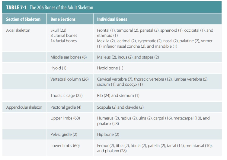
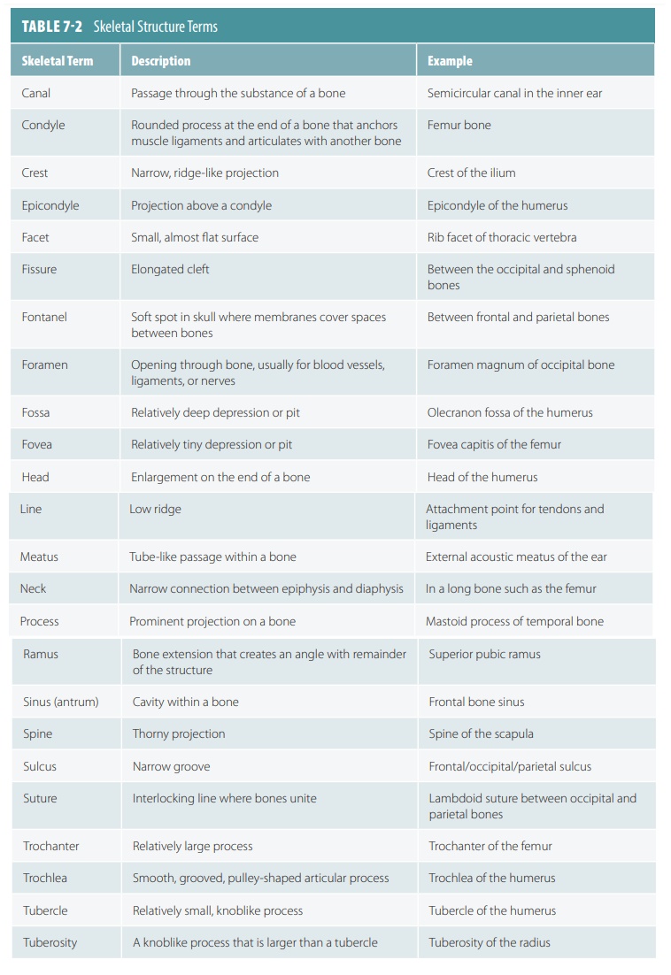
transverse arch. The bones of the adult skeleton are listed in TABLE 7-1 and the
terms describing skeletal structures are listed in TABLE
7-2.
1. Which
bones are involved in the formation of the hip bone?
2. List
the bones of the lower limbs.
3. Which
bone is the strongest in the human body?
4. Which
arches of the feet support the weight of the body?