Pectoral Girdle - Appendicular Skeleton
| Home | | Anatomy and Physiology | | Anatomy and Physiology Health Education (APHE) |Chapter: Anatomy and Physiology for Health Professionals: Support and Movement: Bone Tissues and the Skeletal System
1. Which bone of the upper limbs contains the olecranon fossa? 2. Which bones form the elbow joint? 3. Which bone of the upper limbs contains a styloid process?
Skeletal Organization
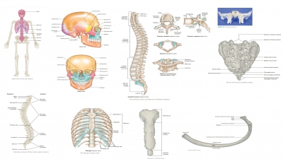
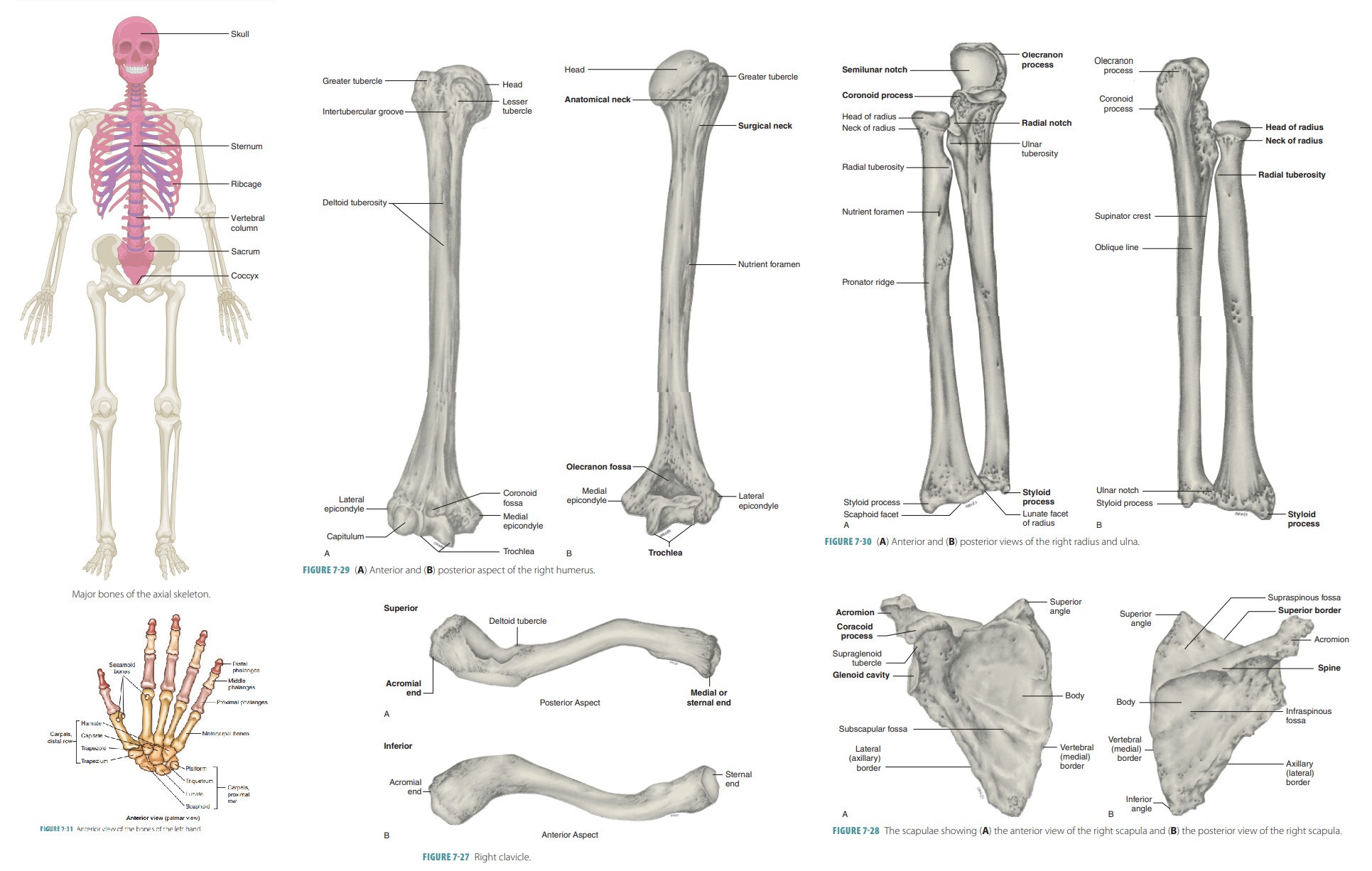
The skeleton is divided into two major portions: the axial skeleton (FIGURE 7-16 ) and the appendicular skeleton. Including those of the middle ear, there are 206 bones in the human body.
Pectoral
Girdle
Appendicular
Skeleton
The appendicular skeleton
contains the upper and lower limb bones and the bones anchoring the limbs to
the axial skeleton. The appendicular skeleton includes the pectoral girdle,
upper limbs, pelvic girdle, and lower limbs.
Pectoral
Girdle
Also known as the shoulder girdle, the pectoral girdle is made
up of a clavicle (collarbone) and a scapula (shoulder blade) on each side of the body. These struc-tures aid in the
movements of the arms. The pectoral girdle is actually an incomplete ring that
opens in the back between the scapulae. It connects the upper limb bones to the
axial skeleton. The sternum separates the bones of the pectoral girdle in the front.
The pectoral girdle supports the upper limbs and is where the mus-cles that
move the upper limbs attach.
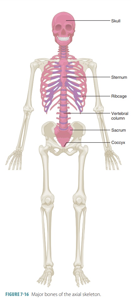
Clavicles
The clavicles or collarbones are shaped like rods with an elongated
S-shape (FIGURE 7-27). They are located at the base of the neck, running horizontally
between the manubrium and the scapulae. These bones brace the scapulae to hold
the shoulders and arms in place and provide muscle attachment points for the
upper limbs, chest, and back. Each clavicle curves laterally and poste-riorly
from its pyramidal sternal end to
form a smooth posterior curve. The flat acromial
end is wider than the sternal end. The clavicles can be easily felt as they
extend horizontally, crossing the upper thorax. The medial two-thirds of each
clavicle are convex anteriorly, while the lateral third is concave anteriorly.
The superior surfaces are mostly smooth, but interior surfaces have ridges and
grooves due to ligaments and action of attached muscles ligament that connects
the clavicle to the scapula is anchored by the trapezoid line and conoid
tubercle.
Fracture of a clavicle causes the
entire shoul-der region to collapse medially. These bones are not as strong as
many other bones in the body. Because of the curves in a clavicle, it often
fractures outward (anteriorly). If it collapses inward (posteriorly),
splin-ters from the bone could damage the subclavian artery, which is just deep
to the clavicle. The clavicles are highly sensitive to muscle action and become
stronger and larger in individuals who use the shoulders and arms during
athletics or labor.
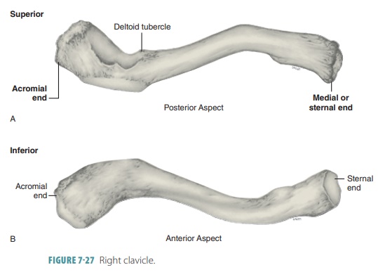
Scapulae
The scapulae or shoulder blades are somewhat triangular bones on
either side of the upper back (FIGURE 7-28). They lie on the dorsal surface of the rib cage, between the second
and seventh ribs. There are three borders in each scapula. The superior border is the sharpest and
short of these. The vertebral (medial)
border is parallel to the vertebral
column. The axillary (lateral) border is
thick, located near the armpit, and extends
superiorly in the small, shallow fossa known as the glenoid
cavity, which articulates with the head of the humerus bone to form the
shoulder joint. Each scapula has three angles,
with the superior scap-ular border meeting the medial border at the superior angle, and meeting the lateral border at the lateral angle. The inferior
angle is the point where the medial and
lateral borders join. The inferior angle has a lot of movement when the arm is
lowered or raised, and is used when scapular movements are assessed.
Each scapula is divided by a
spine that leads to an acromion
process and a coracoid process. The
acromion process provides muscle attachments for the upper limbs and chest and
is continuous with the scapular
spine, a ridge crossing
the posterior scapu-lar surface and ending at the medial border. Where the
acromion process articulates with the acromial end of the clavicle, the acromioclavicular joint is formed.
The coracoid process provides similar attachments and projects from the superior scapular border. It helps anchor the arm’s biceps muscle and is bounded medially by a nerve passage called the suprascapular notch. The coracoid process is bound laterally by the glenoid cavity. The costal (anterior) surface of the scapula is concave, and its posterior surface is where the prominent scapular spine can be felt through the skin. A depression on each scapula’s anterior surface is known as the subscapular fossa. The area superior to the scapular spine is the supraspinous fossa, whereas the area inferior to it is the infraspinous fossa.
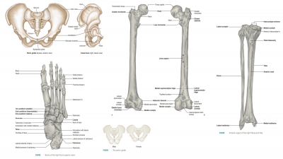
Upper Limbs
The bones of the upper limbs include the arms, forearms, and hands. They
provide muscle attachments and function to move limb parts. The bones of the
upper limbs are:
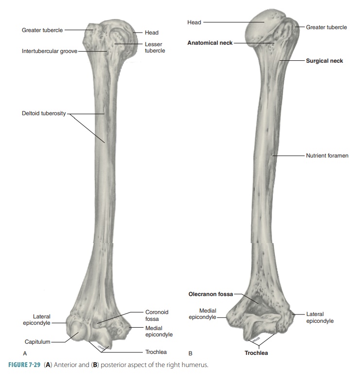
■■ Humerus: This is the upper arm bone, which
extends from the scapula to the elbow. It has a smooth upper head that fits
into the glenoid cavity, with two tubercles providing muscle attachment points
(FIGURE 7-29). The lower portion of the humerus has two smooth condyles that
articulate with the ulna and radius. The narrow depression in this area of the
humerus that separates it from the greater and lesser tubercles s the
anatomical neck. Structures called medial nd lateral epicondyles attach to the
muscles and ligaments of the elbow. The olecranon fossa is a depression on the
posterior surface of the humerus that receives an ulnar olecranon process when
the upper limb straightens at the elbow. The deltoid tuberosity is the large
and rough elevation on the lateral surface of the shaft of the humerus, to
which the deltoid muscle attaches.
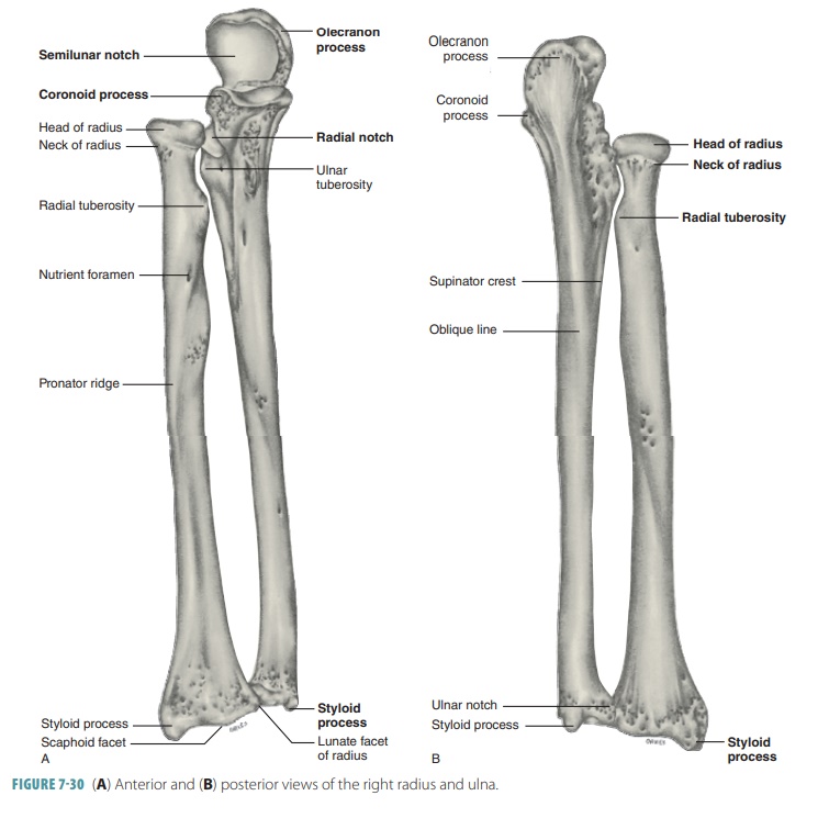
■■ Forearm: The skeleton of the forearm or antebrachium is formed by the
two parallel long bones known as the radius and ulna. In most individuals, these bones can be easily felt. Their proximal ends articulate with the
humerus while their distal ends form joints with the wrist bones. The radius
and ulna articulate with each other proximally and distally at small radioulnar joints. These bones are connected all along their lengths by a
flexible, flat ligament called the interosseous
membrane. The radius lies laterally,
toward the thumb side while the ulna lies medially when in the anatomi-cal
position. When the forearm is rotated and the palm faces posteriorly (the pronation movement), the distal end of
the radius crosses the ulna, with the two bones forming an “X” shape.
■■ Radius: This is located on the thumb side of the forearm. This bone extends from the elbow to the wrist, crossing over the ulna when the hand is turned. Its upper
end articulates with the humerus and a notch in the ulna. A process called the radial tuberosity
serves as an attachment for the biceps brachii muscle. The
distal end of the radius has a styloid
process providing ligament attachments to the wrist. Fracture of the lower end of the radius is called Colles fracture. It is a common injury, con-sisting of fracture of the distal
radial metaphyseal region. Colles fracture is often seen in people who play
high-impact sports or in elderly people who fall onto their hands.
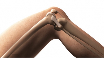
■■ Ulna: Longer than the radius, the ulna overlaps the end of the
humerus and has a trochlear
notch at its proximal end
that articulates with the humerus (FIGURE 7-30). The distal end of the ulna has a head that articulates with the notch of the
radius. The olecranon process and coro-noid process,
located on each side of this notch, provide
attachments for muscles. A disc of fibro-cartilage joins the triquetrum bone of
the wrist.
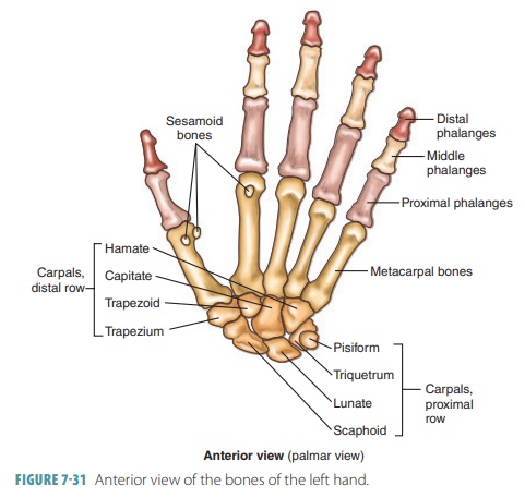
■■ Hand: The hand is the part of the upper limb that consists of the wrist, palm, and fingers. The wrist contains
eight bones called carpals, in two rows of four bones each. They are the scaphoid, lunate,
triquetrum, pisiform, trape-zium, trapezoid, capitate, and hamate bones.
This mass (the carpus)
articulates with radius, ulna, and metacarpal bones. Five bones called metacarpals form the palm (metacarpus)
of the hand. The rounded ends of these
bones form the knuckles and are numbered from one to five, beginning with the
thumb. The metacar-pals articulate with the carpals and phalanges (finger bones). Each finger except the thumb (pollex) has three phalanges
(a proximal, mid-dle, and distal phalanx) . The thumb has only two phalanges
because it lacks a middle phalanx (FIGURE 7-31).
1. Which
bone of the upper limbs contains the olecranon fossa?
2. Which
bones form the elbow joint?
3. Which
bone of the upper limbs contains a styloid process?