Cell Cycle: Cell Division and Cytoplasmic Division
| Home | | Anatomy and Physiology | | Anatomy and Physiology Health Education (APHE) |Chapter: Anatomy and Physiology for Health Professionals: Levels of Organization : Cells
1. Explain the steps in cell division. 2. Why must division of DNA during mitosis be precise? 3. Describe gametogenesis. 4. Compare mitosis and meiosis.
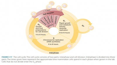
Cell Cycle
The life cycle of each cell is
regulated via stimulation from hormones or growth factors. Disruption of the
cycle can affect the health of the body. Most human cells divide from 40 to 60
times before they die. The life cycle of a cell includes the following steps:
■■ Interphase: The cell obtains
nutrients to grow andduplicate. This is actually the period from cell for
mation to cell division. This step may be betterunderstood as being a metabolic
or growth phase.
■■ Cell division (mitosis): The
nucleus divides.
■■ Cytoplasmic division
(cytokinesis): The cytoplasmdivides.
■■ Differentiation: The cell becomes specialized.
Cell Division
and Cytoplasmic Division
The two types of cell division
are meiosis and mitosis/cytokinesis. Meiosis is part of gametogenesis (the
for-mation of egg or sperm cells depending on gender). Meiosis reduces by half
the number of chromo-somes, from 46 to 23, in eggs and sperm, so when they
unite the fertilized egg will have the proper total of 46 chromosomes. Mitosis
is characteristic of the somatic cells. Two new daughter cells result from cell
division, receiving the same number of chromosomes present in the parent cell.
Mitosis
Mitosis occurs in the somatic cells, but not in everymature cell such
as cardiac muscle and nerve cells. Cell numbers increase via this process, in
which cell nuclei divide. In cytokinesis,
the cytoplasm of a cell divides. All cells, except egg and sperm cells, can be
divided by mitosis. When the nucleus divides, it must be pre-cise so an
accurate copy of the DNA information can be made by the new cell. Connective
tissue and liver cells are examples of cells that divide as needed to heal
injury or to replace lost or damaged cells. Cells of the digestive tract and
bone marrow divide continually. Overall, the rate of cell division is
controlled by the body so that excess cells are not produced.
Growth-promoting substances
called growth factors are secreted by nearby cells. They bind totarget cell
membrane receptors, activating them. The activated receptors transmit signals
that cause the cell to divide, assisted by genes. The effects of genes include
promotion of cell growth by producing cell surface receptors to which growth
factors attach. Other genes create signals that suppress cell growth and
division. Normal cells divide enough to be func-tional and to replenish
cellular loss from aging or injury. Normal cells cannot continue to divide
for-ever, but have a limited number of cell divisions before they die. The
process of neoplasia or dysreg-ulated cell growth is linked to defective
regulation of cell division, and may lead to cancer.
A cell’s DNA chains are
duplicated, forming new chromosomal material known as the “S” phase. The
chromosomes and their duplicates are located next to each other. The two
members of the pair are known as chromatids. In mitosis, the chromatidsseparate. During cell division, when
chromosomes condense, each of them actually consists of two sep-arate
chromosomes that are partially joined where their spindle
fibers attach. The term “chromatid”
describes these still joined chromosomes. Once they separate, they are again
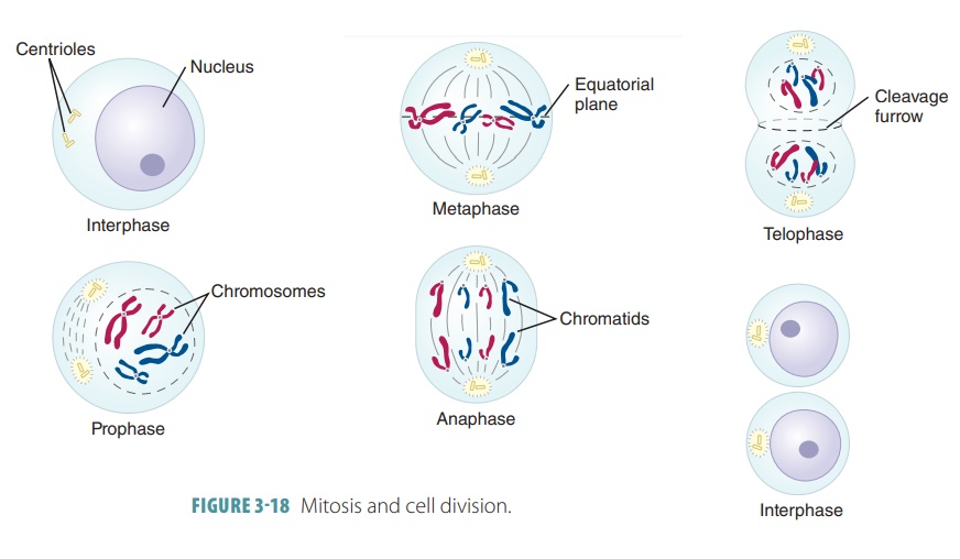
called chromosomes. There are several stages of mitosis, including prophase,metaphase, anaphase, andtelophase.
Prophase
In prophase, each chromosome becomes thicker and shorter (FIGURE 3-18). The centrioles move to opposite poles of the
cell. They form the mitotic spindle,
consist-ing of small fibers that radiate in many directions and form the
centrioles. Some spindle fibers attach to the chromatids. The nuclear membrane
breaks down near the end of prophase.
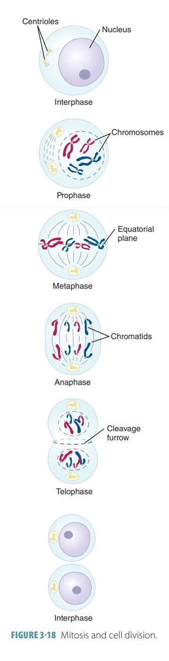
Metaphase
In metaphase, the chromosomes line up near the mid-dle portion (the equator of the cell) between the
cen-trioles. The chromatids are partially separated but still joined, with
spindle fibers attached to them at a con-stricted section called the centromere.
Anaphase
In anaphase, the chromatids of each chromosome are pulled apart to
become individual homologous chro-mosomes. Pulled by the spindle fibers, they
move toward opposite ends (poles) of the cell.
Telophase
In telophase, the spindle fibers disappear and the chromosomes
lengthen and unwind. A nuclear enve-lope forms around them and nucleoli appear
in each newly formed nucleus. The nuclear membranes of the two daughter cells
reform. The cytoplasm divides to form two daughter cells that are exact
duplicates of the parent cell.
Cytoplasmic division
(cytokinesis) actually begins during anaphase when the cell membrane constricts
down the middle portion of the cell. However, this continuous process is
completed through telophase to divide the cytoplasm. The two newly formed
nuclei are then separated, and nearly half of the organelles are distributed
into each new cell.
Gametogenesis
The gonads, consisting of the testes and ovaries, con-tain precursor cells known
as germ cells. These can develop into
mature sperm or ova. When mature, germcells are called gametes. The
process in which they form is known as gametogenesis.
Two similar processes occur in males and females: sperm develop during spermatogenesis and ova develop during oogenesis.
Spermatogenesis
In the testicular tubules,
precursor cells are called spermatogonia. They each contain 46 chromo-somes, and divide via mitosis
forming primary spermatocytes . Like precursor cells, spermatocytes also contain 46
chromosomes. Primary spermato-cytes then divide by meiosis. In the first
division, each primary spermatocyte forms two secondary sper-matocytes, with
each containing 23 chromosomes. The secondary spermatocytes complete the second
meiotic division forming two spermatids . These also contain 23 chromosomes and eventually mature
to become sperm. Spermatogenesis takes about 2 months. Sperm are continually
produced after the male reaches sexual maturity.
Oogenesis
Precursor cells of the ova are
known as oogonia. They each contain 46 chromosomes, but divide repeatedly in the fetal
ovaries prior to birth. This forms primary oocytes, also containing 46 chromosomes. A singlelayer of granulosa cells then surround the oocytes. Also called follicular cells, the granulosa cells form the primary follicles. Inside these follicles, the primary oocytes
begin, but do not complete prophase of the first meiotic division during fetal
life. Large numbers of primary follicles are formed, with many degener-ating
during infancy and childhood. As many as 20% of oocytes have chromosome
complement or aneu-ploidydefects.
Even so, about 500,000 primary folliclespersist into adolescence. The loss of
primary follicles continues throughout a female’s reproductive years. In every
reproductive cycle, several oocytes start to mature. Usually, only one oocyte
is ovulated, while the others degenerate. In menopause, only several thousand
oocytes are left. Their numbers decline until there are no more oocytes left in
the postmenopausal female’s ovaries.
The ovaries and their primary
follicles are inactive until puberty. The cyclic ovulation starts, influenced
by the pituitary gonadotrophic hormones, which include follicle-stimulating hormone (FSH) and luteinizing
hormone (LH). In every menstrual cycle, a number of primary follicles start to
grow. Usually, only one follicle reaches full maturity and is ovulated. When
the oocyte is discharged, the first meiotic division is completed. Two daughter
cells develop of different sizes. One daughter cell receives half of the
chromo-somes, which is one member of each homologous pair. It also receives
nearly all of the cytoplasm. This daughter cell is called secondary oocyte and
contains 23 chromosomes. The other daughter cell receives theremaining 23
chromosomes, but nearly no cytoplasm. It is called the first polar body and is
eventually dis-carded. The new secondary oocyte quickly begins its second
meiotic division. This leads to the formation of a mature ovum and a second
polar body. Each of these contains 23 chromosomes. The meiotic division is not
completed unless the ovum is fertilized.
Meiosis
In meiosis, cell division reduces
the amount of chro-mosomes by half. There is a mixing of genetic mate-rial
between homologous chromosomes. This is a type of recombination process, which is referred to as crossing
over. There are two separate meiotic
divisions:
■■ First meiotic division: Like
mitosis, every chro-mosome is duplicated prior to cell division. Two chromatids
are formed. In prophase, each pair of homologous chromosomes lie next to each
other over their entire length called a synapse.
Some interchange of segments occurs called a crossover, a characteristic feature of meiosis. In females, the two
X chromosomes synapse exactly like autosomes. In males, the X and Y chromosomes
synapse end to end, with no segments being exchanged. Crossing over is faster
in females than in males. In metaphase, paired chromosomes are arranged in a
plane in the middle of the cell. In anaphase, the chromosomes separate and move
to opposite poles in the cell. The chromosomes each consist of two chromatids
that do not yet sepa-rate. In telophase, two new daughter cells form, each
containing only one member of each pair of homologous chromosomes. Therefore,
the chro-mosomes in each daughter cell are reduced by one-half. These
chromosomes are different from those of the parent cell, due to the interchange
of genetic material during synapse.
■■ Second meiotic division:
Similar to a mitotic divi-sion, the two chromatids making up each chro-mosome
separate. Two new daughter cells are formed. Each of them contains half of the
normal number of chromosomes.
1. Explain
the steps in cell division.
2. Why
must division of DNA during mitosis be precise?
3. Describe
gametogenesis.
4. Compare mitosis and meiosis.