Movement of Digestive Materials
| Home | | Anatomy and Physiology | | Anatomy and Physiology Health Education (APHE) |Chapter: Anatomy and Physiology for Health Professionals: Digestive System
There are two basic types of motor functions in the alimentary canal: mixing movements and propelling movements.
Movement
of Digestive Materials
There are two basic types of motor functions in the
alimentary canal: mixing movements and propelling movements. When smooth
muscles contract rhythmically, mixing occurs. Waves of contractions mix food with
digestive juices. In the small intestine, mixing movements are aided by
segmentation, involving alter-nating contraction and relaxation of smooth
muscle in nonadjacent segments. Materials are not propelled along the tract in
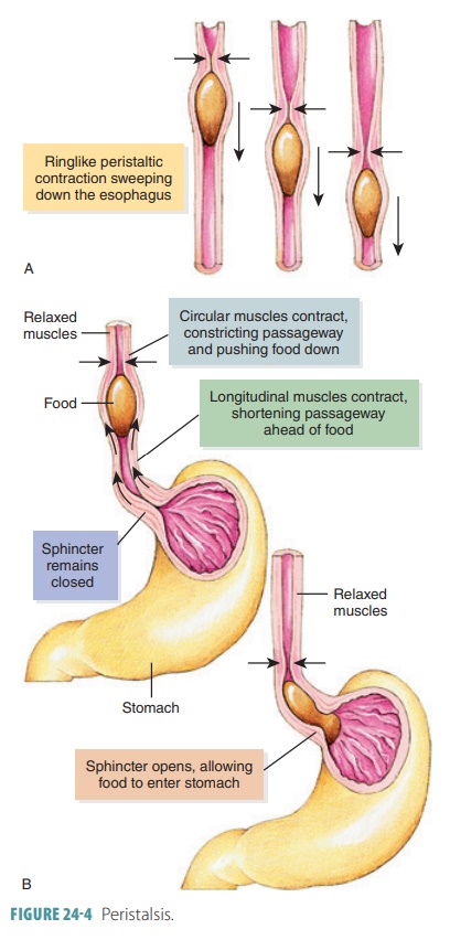
one direction because segmentation follows no set pattern. Peristalsis consists
of the propel-ling, wave-like movements of the tube (FIGURE 24-4). Contraction appears in the wall
of the tube in a “ring,” whereas the muscular wall immediately ahead of the
ring relaxes. The peristaltic wave moves along, pushing contents of the tube
toward the anus.
Blood Supply
The arteries that branch from the abdominal aorta, serving
the digestive organs and hepatic portal
circu-lation, make up the splanchnic circulation. Usually, one-fourth of the cardiac output supplies these arter-ies, which
include branches of the celiac trunk serving the liver, spleen, and stomach.
They also include the mesenteric arteries serving the small and large intes
tines. After a meal, the amount of blood needed from the heart increases.
Venous blood that is rich in nutri-ents is collected by the hepatic portal
circulation from the digestive viscera. It is then transported to the liver.
Enteric Nervous System
The enteric
nervous system is the nerve supply within the alimentary canal. Its
semi-autonomous enteric
neurons communicate with each other, reg-ulating
digestive activities. They make up the bulk of the two primary intrinsic nerve plexuses, which are
ganglia that are interconnected by unmyelinated fiber tracts. These submucosal and myenteric nerve plexuses are within the alimentary canal walls.
They are inter-connected throughout the GI tract and regulate the activities of
digestion all along the tract.
The submucosal nerve plexus is within the submucosa. The myenteric nerve plexus is like a sandwich, between the circular and longitudinal muscle layers that form the muscularis externa. The enteric neurons are the major nerve supply of the GI tract walls. They control motility, which is the motion of substances through the tract. Within the enteric nervous system, there are two types of reflex arcs:
■■ Short
reflexes: These are completely under enteric nervous system plexus control.
They respond to GI tract stimuli. Segmentation and peristalsis is mostly under
automatic control via the pacemaker cells and reflex arcs between the enteric
neurons in various organs.
■■ Long
reflexes: These are controlled by CNS integration centers and extrinsic
autonomic nerves. The enteric nervous system communicates with the CNS over
afferent visceral fibers, receiving sympathetic and parasympathetic branches or
motor fibers, of the autonomic nervous system that enter intestinal walls and
synapse with intrinsic plexus neurons. Long reflexes may be started via stimuli
from inside or outside the GI tract. For these reflexes, the enteric nervous
system handles autonomic impulses to allow extrinsic controls to affect
digestion. Basically, parasympathetic impulses increase digestive activities
while sym-pathetic impulses reduce them.
1. Describe the organs of the alimentary canal from beginning
to end.
2. Describe the wall of the alimentary canal.
3. Define peristalsis.
4. Describe the functions of the mucosa (mucous membrane) of
the alimentary canal.
5. Differentiate between the submucosa, muscularis externa,
serosa, and adventitia.
6. Explain how the splanchnic circulation works.

