Teeth
| Home | | Anatomy and Physiology | | Anatomy and Physiology Health Education (APHE) |Chapter: Anatomy and Physiology for Health Professionals: Digestive System
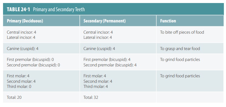
Humans have two sets of teeth that form during their lifetimes. The primary teeth, also called deciduous teeth, erupt through the gums between the ages of six months and four years. These are also known as the baby teeth or milk teeth.

Teeth
Humans have two sets of teeth that form during their lifetimes. The primary teeth, also called deciduous teeth,
erupt through the gums between the ages of six months and four years. These are also
known as the baby
teeth or milk
teeth. There are a total of 20 deciduous teeth, 10 in each jaw, which is
made up by the mandible and maxilla. Each tooth is clinically described as a dentition. The lower central
incisors are the first teeth to appear, usually at the age of six months. In
one to two-month intervals, additional pairs of teeth erupt. In most cases, all
20 primary teeth have emerged by the age of two years. They are eventu-ally
shed, usually in the order they appeared.
The secondary teeth
or permanent teeth push the
primary teeth out of their sockets called alve-oli,
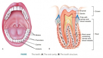
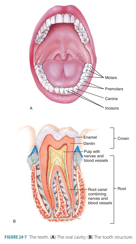
and usually consist of 32 teeth (16 in each jaw; FIGURE
24 -7A). They lie more deeply than the primary teeth and absorb
the roots of the primary teeth from below. The secondary teeth usually begin to
appear at six years of age, but the final teeth, the third molars, may not
appear until between ages 17 and 25. The third molars are commonly referred to
as wisdom teeth. However, in some individuals, wisdom teeth either never emerge or are totally absent.
The primary and deciduous teeth are summarized in TABLE 24-1.
Teeth are classified by their shapes and functions, and
include incisors, canines, premolars, and molars. Incisors are shaped like chisels and are ideal for
cut-ting food (chopping) or for slicing off pieces of food. The canines, also known as the cuspids or eyeteeth, are cone-shaped and resemble fangs. The cuspids are
pointed teeth that are adapted for tearing and shred-ding. The teeth best used
for crushing or grinding are the premolars
or bicuspids and molars. Both have broad crowns and rounded tips called
cusps. Extra grinding ability is
found with the molars because they have four to five cusps. Great crushing
forces are developed as the upper and lower molars lock together during
chewing.
Tooth Structure
Each tooth consists of a crown that projects beyond the gum, also called the gingiva, and a root that
is anchored to the jaw’s alveolar process. The alveolus is the pink ridge that
surrounds the bases of the teeth. The crown of each tooth surrounds it tightly,
like a collar. A gingival sulcus,
which is a shallow groove, surrounds the neck of each tooth. Its mucosa is thin
and not tightly connected to the periosteum. The epithelium is attached to the
tooth at the base of the sulcus, preventing bacte-rial access to the lamina
propria of the gingiva and to the cementum of the root, which is relatively
soft. These attachments are strengthened and the epithelial cells stimulated
when you brush and massage your gums.
The crown of each tooth is covered in glossy, white enamel that mainly consists of calcium salts. The enamel is the
hardest substance in the body (FIGURE
24- 7B), even harder than bone. However, it is brittle and similar
to the substance called ceramic. It
is only about as thick as a dime and bears the force of chewing directly on it.
Enamel contains force-resisting columns of dense hydroxyapatite crystals,
which are minerals aligned perpendicular to the surface of each tooth. When a
tooth erupts, the cells producing the enamel degenerate. Therefore, when
damaged by abrasion or injury, enamel is not replaced, and it wears away with
age. This requires artificial fillings to replace the lost areas of enamel.
The neck of the
tooth is constricted, connecting the crown and the root. The root is enclosed in thin, bone-like cementum surrounded by a periodontal ligament, meaning the root is actually embedded in the jawbone. The cementum is a calcified
connective tissue. The periodontal ligament contains blood vessels and nerves
along with thick collagenous fibers passing between the cementum and bone of
the alveolar pro-cesses. The periodontal ligament anchors the tooth in its alveolus, and a fibrous joint or gomphosis is formed.
One root is found in each of the canines, incisors, and
premolars. However, the first upper premolars usually have two roots. There are
three roots to the first two upper molars, but the corresponding lower molars
only have two. Although the roots of the third molars vary, a fused single root
is most commonly seen.
The bulk of a tooth below the enamel is the dentin, which is harder than bone but similar in structure. It surrounds the tooth’s central pulp cavity that contains blood vessels, connective tissue, and nerves. Collectively, these components are called pulp. Blood vessels and nerves reach the pulp cav-ity through root canals extending into the root. The dentin is rich in proteins, acting as a shock absorber when chewing and biting. It contains unique radial striations known as dentinal tubules . Dentin, cemen-tum, and enamel are calcified. They differ from bone in that they are avascular. Also, dentin and cemen-tum contain collagen, whereas enamel does not and is almost all mineral in content. The pulp supplies nutri-ents to the tissues of the tooth and provides sensation. At the point where the pulp cavity extends into the tooth root, it becomes the root canal. The proximal end of each root canal has an apical foramen, which allows blood vessels, nerves, and other structures to enter into the pulp cavity. The superior and inferior alveolar nerves, which are branches of the trigeminal nerve, serve the teeth. Blood is supplied by branches of the maxillary artery called the superior and infe-rior alveolar arteries.
Tooth Disease
When bacteria eventually demineralize enamel and dentin, cavities or dental caries result. The decay starts when a
film of bacteria, sugar, and various debris adheres to the teeth. This is known
asdental plaque. Acids
are produced, which dissolve the calcium salts in the teeth. Bacterial enzymes
can then easily digest the rest of the organic teeth matrices. Plaque can be
removed by frequent brushing and daily flossing.
Gum Disease
When plaque is not removed from the gums, it calci-fies,
forming a calculus, which is also
known as tartar. This condition is
more serious than tooth decay. The extremely hard calculi disrupt the seals
between the gums and teeth. The sulci are deepened and the gums may become
infected by pathogenic anaerobic bacte-ria, a condition called gingivitis. The gums become red, swollen,
sore, and often bleed. If the calculi are removed, gingivitis is reversible. If
not, inflamed pock-ets of infected tissue develop. Immune system cells and
neutrophils then attack the pathogens as well as the gum tissues. Deep pockets
develop around the teeth, the periodontal ligament is destroyed, and bone is
dis-solved by activated osteoclasts. This condition, known as periodontitis (periodontal disease), affects nearly 95% of people over the age of
35. In adults, periodontal disease causes between 80% and 90% of tooth loss.
However, intervention is still possible. The teeth are
scraped, the infected pockets cleaned, and the gums are cut to shrink the size
of the pockets. Anti- inflammatory and antibiotic therapies are initiated. The
surrounding tissues can then reattach to the teeth and bone. Home regimens
follow, including consistent brushing, flossing, and rinsing with hydrogen
peroxide mixtures. It is also believed that untreated periodon-tal disease may
be linked to heart disease and stroke. This is because the existing, chronic
inflammation promotes atherosclerotic plaques, and bacteria that enter the
blood from the infected gums result in clots to form, clogging cerebral and
coronary arteries. Peri-odontal disease is commonly linked to diabetes
melli-tus, smoking, and piercings of the tongue or lips.
1. Describe the time periods involved in the eruption of the
primary and secondary teeth.
2. Explain the tooth substance that is harder than bone yet
can become permanently eroded.
3. Why is gum disease more serious than tooth disease?