Spinal Cord
| Home | | Anatomy and Physiology | | Anatomy and Physiology Health Education (APHE) |Chapter: Anatomy and Physiology for Health Professionals: Central Nervous System
The spinal cord is a thin column of nerves leading from the brain to the vertebral canal.

Spinal Cord
The spinal
cord is a thin column of nerves
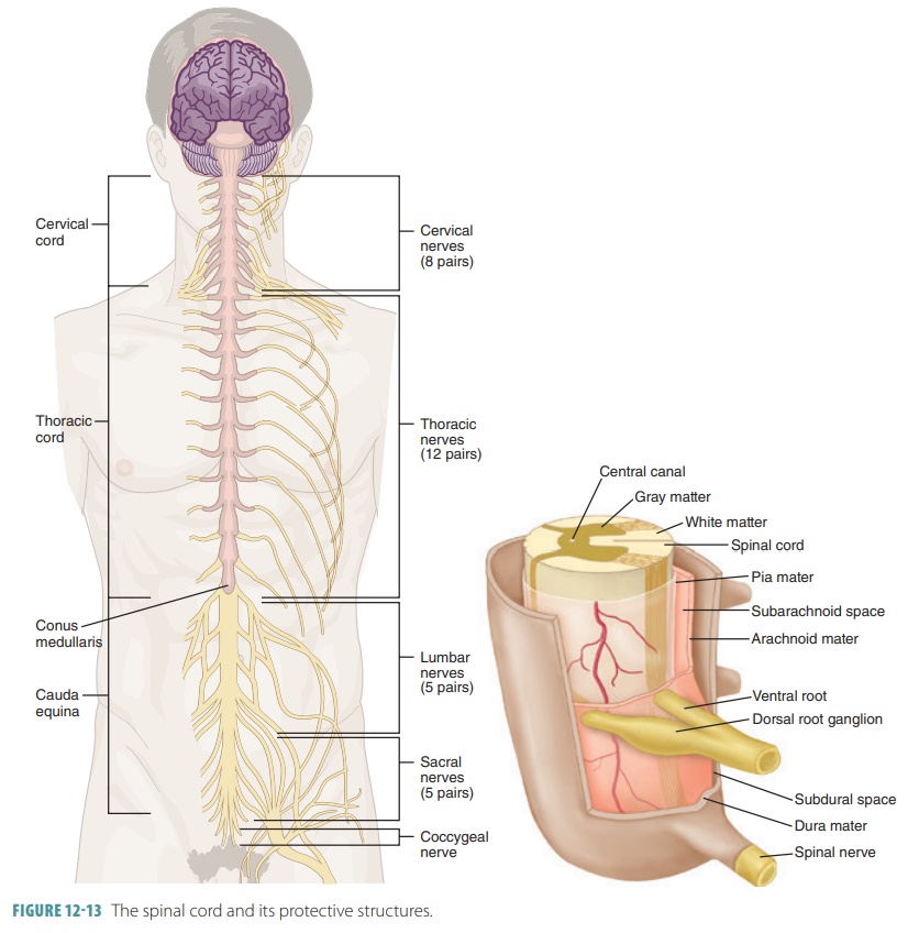
leading from the brain to the vertebral canal. It starts at the point where
nervous tissue exits the cranial cavity near the foramen magnum and eventually
tapers off to terminate near the point where the first and second lumbar are
located (FIGURE 12-13). The spinal cord is approximately 42 cm in length and 1.8 cm thick. It
appears as a shiny white structure, protected by bone, meninges, and CSF. The
spinal cord provides two ways of communication, to and from the brain, and
contains the spinal reflex centers. The spinal cord is continuous throughout
its length, with slight internal structure changes. The spinal cord is divided
into right and left halves by a deep anterior median fissure and a shallow
posterior median sulcus. It is slightly flat from front to back.
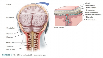
The spinal meninges are
continuous with those of the brain. In the spinal cord, the dura mater has a single layer and is
not attached to the vertebral col-umn. The inner surface of the dura mater
contacts the outer surface of the arachnoid
mater, which is the middle meningeal layer of the spine. An epidural space exists between the
dura mater and vertebrae, which is padded by fat and a vein network. The CSF fills the
subarachnoid space, which lies
between the arachnoid and pia mater meninges. The dural and arachnoid membranes
extend inferiorly to the S2 level, which is far below the end of the
spinal cord that ends between the L1 and L2 levels. The
subarach-noid space inside the meningeal sac inferior to the lumbar region is
an excellent spot for the removal of CSF. This procedure is called a lumbar puncture or spinal tap.
The spinal cord terminates
inferiorly in a tapered, cone-shaped structure called the conus medullaris. A
fibrous extension of the conus medullaris called the filum terminale extends
inferiorly to the coccyx to anchor the spinal cord. Sawtooth- shaped sections
of pia mater are called denticulate
ligaments and bind the spinal cord to the
dura mater meninx for its entire length. Components of the filum terminale
blend with a dense cord of collagen fibers continu-ous with the spinal dura
mater to form the coccygeal ligament. The spinal cord is only about
as wide as a human thumb over most of
its length. It has prom-inent enlargements at points where the nerves arise
that serve the upper and lower limbs. These are called the cervical and lumbar
enlargements.
Cross-Section of the Spinal Cord
The inner core of the spinal cord
is made of gray mat-ter surrounded by white matter. Motor fibers pass out of
portions of the gray matter through spinal
nerves to skeletal muscles; however, most of the gray mat-ter neurons are
interneurons. Each segment of the spinal cord is designated by its paired
spinal nerves. Each spinal nerve emerges from the vertebral column superiorly
to its related vertebra via the intervertebral foramen. Each nerve travels to
the body region that it specifically serves.
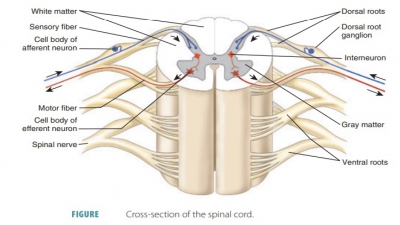
The sensory fibers that enter the
spinal cord usu-ally end on interneurons, which receive input from the sensory neurons.
Impulses may be transmitted by the interneurons to adjacent multipolar motor
neurons. The axons of these motor neurons leave in the spinal nerve’s ventral root and nerve impulses are
transmitted to muscles and glands. The arrangement of these neurons enables
information to enter and leave the spinal cord very quickly. The spinal cord
conducts not only nerve impulses, but also spinal
reflexes.

The gray matter of the spinal
cord appears as a butterfly shape, also described as similar to the letter “H,”
within the spinal cord’s white matter. Major nerve pathways called nerve tracts are made
up of long bundles of myelinated nerve fibers. A horizontal bar of gray matter
in the very middle of the spinal cord surrounds its central canal and contains
CSF. This is known as the gray
commissure. Its two dorsal gray matter
projections are called the dorsal
(posterior) horns, whereas the ventral
pair is called the ventral
(anterior) horns (FIGURE 12-14). They run the entire length of the spinal cord. The dorsal horns contain somatic
and visceral sensory inputs, which
receive and relay sensory information for peripheral receptors. An additional
pair of gray matter columns called the lateral
horns exists in the
thoracic and superior lum-bar segments (sympathetic neurons).
All neurons that have cell bodies in the gray matter of the spinal cord are multipolar. Interneurons com-pletely make up the dorsal horns, whereas the ventral horns have mostly somatic motor neurons with lower amounts of interneurons. The motor neurons send their axons out to the skeletal muscles, which are their effector organs, via ventral rootlets. These rootlets fuse together, becoming the spinal cord’s ventral roots. Sensory and motor roots are bound together into one spinal nerve. This occurs distal to each dorsal root ganglion.
Cell bodies of autonomic
(sympathetic division) motor neurons, which serve visceral organs, mostly make
up the lateral horns. Their axons leave the spinal cord with the ventral root
alongside those from the somatic motor neurons. Ventral roots serve both PNS
motor divisions because they contain somatic and autonomic efferent fibers.
The dorsal
roots are formed by afferent fibers,
which carry impulses from peripheral sensory recep-tors. They fan out as the dorsal rootlets before entering the
spinal cord. Associated sensory neuron cell bodies lie in an enlarged region of
each dorsal root, which is known as the dorsal
root ganglion (spinal ganglion).
The white matter of the spinal
cord is made up of myelinated and nonmyelinated nerve fibers. These allow
communication between sections of the spi-nal cord and between the spinal cord
and brain. The nerve fiber tracts run in three different directions. The tracts
that carry information to the brain are called ascending tracts and the tracts that carry infor-mation to the
muscles and glands are called descend-ing
tracts. The ascending tracts run up to higher centers for sensory input and the descending tracts run down from
the brain to the spinal cord or inside the spinal cord to its lower levels for
motor output.
The transverse tracts run across
the spinal cord from one side to the other with commissural fibers. Most white
matter is made up of ascending and descend-ing tracts.
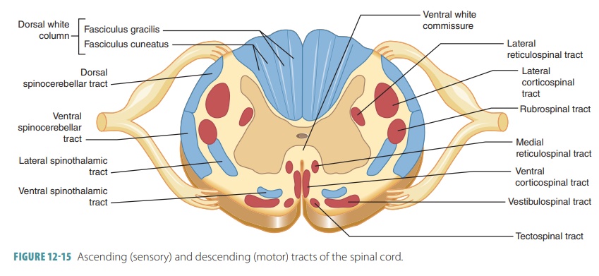
There are three white matter
columns called funiculi on each side of the spinal cord. They are named by their
positions as the dorsal (posterior) funiculi, the lateral funiculi, and the
ventral (anterior) funiculi. Each spinal tract contains several fiber
tracts made up of axons that have
similar functions and destinations. The names of the spinal tracts describe
their destinations as well as origins (FIGURE
12- 15). The anterior white
columns are interconnected by the anterior
white commissure, which is where axons cross from either side of the spinal cord. The lateral white column is
made up by the white matter between the anterior
and posterior columns on each side.
The roots of the lumbar and sacral spinal nerves angle sharply downward. They travel inferi-orly through the vertebral canal for a long distance, finally reaching their intervertebral foramina. At the inferior end of the vertebral canal is a collection of nerve roots called the cauda equina, since it looks like a horse’s tail.
1. List
the layers of the spinal cord.
2. What
are the two major functions of the spinal cord?
3. Explain
the cervical and lumbar enlargements of the spinal cord.