Structures of Bones
| Home | | Anatomy and Physiology | | Anatomy and Physiology Health Education (APHE) |Chapter: Anatomy and Physiology for Health Professionals: Support and Movement: Bone Tissues and the Skeletal System
Bones are considered organs because they contain various types of tissue.
Structures of
Bones
Bones are considered organs
because they contain various types of tissue. They are not only dominated by
osseous (bony) tissue, but also contain nervous tissue, cartilage, fibrous
connective tissue, and both muscle and epithelial tissues. Nervous tissue is
found in bone nerves, whereas cartilage is found in articular cartilages.
Fibrous connective tissue lines bone cavi-ties, and both muscle and epithelial
tissues are found in the blood vessels of bones.
Gross Anatomy
The external layer of bones is
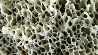
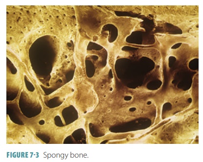
called compact bone, which contains spongy bone that is made up of small flat or needle-like pieces called trabeculae (FIGURE 7-3). It
appears smooth and solid to the naked eye. Open spaces between trabeculae are
filled with red and yellow bone marrow. Spongy bone is made up of open struts and plates, covered by a thin cortex of compact bone. This covering is
also referred to as cortical bone.
Bones described as short,
irregular, or flat are all made up of thin plates of the spongy bone covered by
the compact bone. The plates are covered by connec-tive tissue membranes (the periosteum outside and the endosteum inside). These bones have no
shaft or epiphyses because they are not cylindrical. Inside, they have bone
marrow between their trabeculae, although there is not a well-formed marrow
cavity. Hyaline car-tilage covers the surfaces of these bones where they form
movable joints with nearby bones. The spongy bone in flat bones is called the diploe.
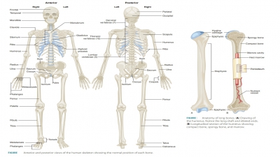
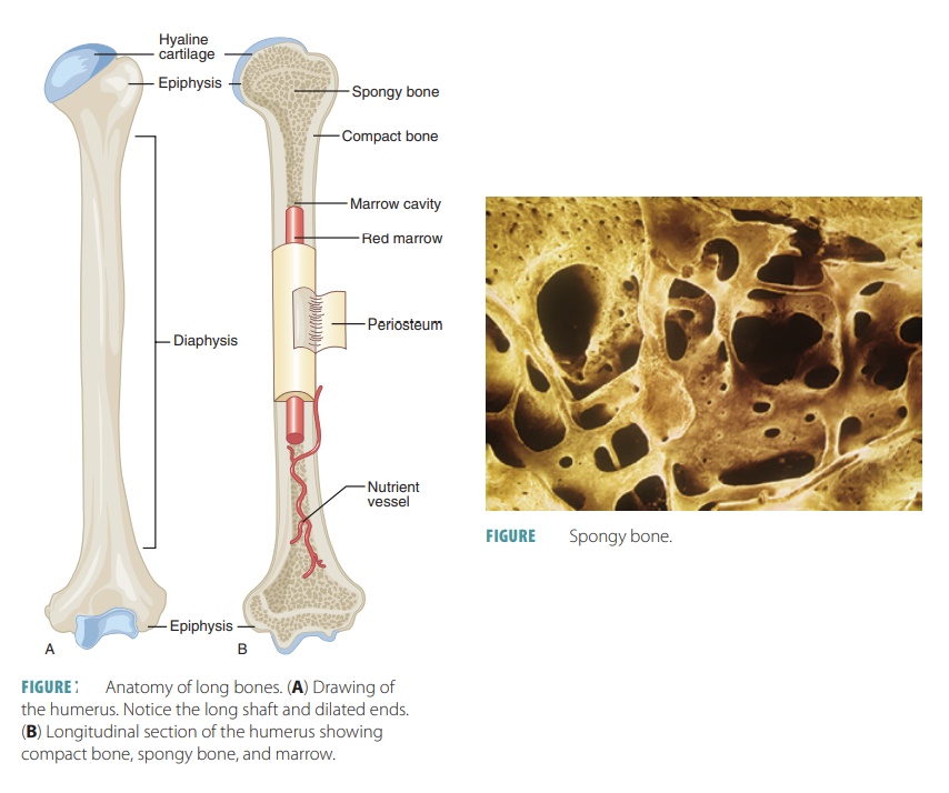
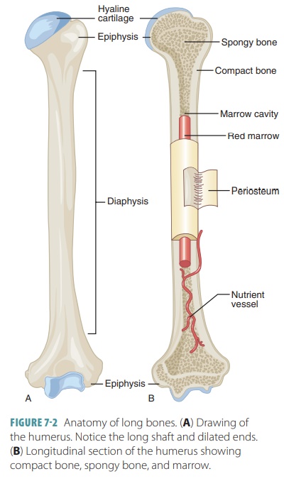
Nearly all long bones have the structure of a shaft, bone ends, and membranes. The shaft is known as the diaphysis. It is a tubular structure forming the long bone axis, made of a thick collar of compact bone sur-rounding a central medullary cavity. This cavity is called the yellow marrow cavity in adults because it contains fat (yellow marrow).
The bone ends are called epiphyses, which
are usually broader than the diaphysis. Inside, they have spongy bone, whereas
their outer shell is made of compact bone. The joint surface of each epiphysis
is covered by a thin layer of hyaline cartilage. This layer cushions opposing
bone ends as they move and absorbs stress. Between the diaphysis and each
epiph-ysis of long bones is an epiphyseal
line, which is a left-over remnant of the epiphyseal
plate. There is also a disc of hyaline
cartilage that develops during child-hood, lengthening the bone. At the point
where the diaphysis and epiphysis meet is a flared portion that is sometimes
referred to as the metaphysis.
The external surface of a bone,
except for the joint surfaces, is covered by a shiny, white, and double-
layered periosteum, a membrane that is richly supplied with blood vessels and nerve fibers.
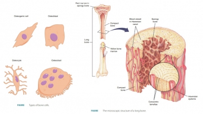
Dense irregular connective tissue makes up the outer fibrous layer. Inside, an osteogenic layer touches the bone
sur-face and is made up mostly of primitive stem cells known as osteogenic
cells. These cells form all bone
cells, except for those that function in bone destruc-tion. The blood vessels
and nerve fibers of the peri-osteum pass through the bone shaft, entering the
marrow cavity through openings known as nutrient
foramina. Groups of collagen fibers,
called perforat-ing fibers or Sharpey’s fibers, extend from the
fibrous layer into the bone matrix
and bind the periosteum to the underlying bone. The periosteum also serves to
anchor tendons and ligaments in areas that are extremely dense. Metaphyseal vessels supply blood to the
diaphyseal surface of the epiphyseal cartilages, where they are replacing bone.
Periosteal vessels provide blood to
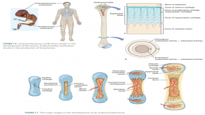
the superficial osteons of the bone shafts. During endochondral bone formation,
they branch to enter the epiphyses and provide blood to the secondary ossification
centers.
The endosteum is a delicate connective tissue membrane covering internal bone
surfaces. In most trabecular cavities of spongy bone (in long bones) and in the
diploe of flat bones are found hematopoietic red marrow. As a result, both these cavities are referred to as red marrow cavities. The endosteum
covers the trabec-ulae of spongy bone and lines canals that pass through
compact bone. The endosteum also contains osteogenic cells, which may
differentiate into various bone cells.
In the newborn, red bone marrow
is found in the medullary cavity of the diaphysis as well as all areas of
spongy bone. In adults, most long bones have a medullary cavity that contains
fat, which extends a good length into the epiphysis. Very little red marrow is
found in an adult’s spongy bone cavities. As a result, red blood cell
production in adult long bones usually occurs only in the heads of the femur
and humerus. There is a greater amount of hematopoietic activity in the red
marrow of the diploe of flat bones (such as the sternum) and certain irregular
bones (such as the hip bones). Yellow marrow in the medullary cavity can convert back to red marrow if there is
significant ane-mia, requiring more red blood cells.
Bone Markings
External surfaces of bones
usually have depressions, projections, and openings. These bone markings are where ligaments, muscles, and tendons attach or
they may occur at joint surfaces. They also may serve as conduits for nerves
and blood vessels. Pro-jections bulge outward from bone surfaces, and include
heads, spines, trochanters, and others. Each of these has its own unique
features. Most bone pro-jections show stresses caused by attached muscles or
are modified surfaces where the bones meet and form articulations.
Depressions and openings in bones
allow for pas-sage of nerves and blood vessels and include:
■■ Fissures: Narrow, slit-like
openings
■■ Foramina: Oval or round
openings through bones
■■ Grooves: Shallow depressions
■■ Notches: Indentations at the
edges of structures
■■ Fossae: Shallow depressions in
bones that often serve as articular surfaces
■■ Meatuses: Passageways that
resemble canals
■■ Sinuses: Cavities inside bones
that are filled with air and lined with mucous membranes
Bone projections that are the
sites where muscles and ligaments attach are:
■■ Crests: Narrow, usually
prominent ridges of bone
■■ Epicondyles: Raised areas on
or above condyles
■■ Lines: Narrow ridges of bone
that are not as prominent as crests
■■ Processes: Bony prominences
■■ Spines: Pointed, sharp, or
slender projections
■■ Trochanters: Extremely large, blunt,
irregular shaped processes that only occur on the femurs
■■ Tubercles: Small rounded
projections or processes
■■ Tuberosities: Large rounded
projections that may be rough
Bone projections that help to
form joints include:
■■ Condyles: Rounded articular
projections
■■ Facets: Smooth, almost flat
articular surfaces
■■ Heads: Bony expansions that
are carried on narrow necks
■■ Rami: Arm-like bars of bone
1. Which
membranes cover the external and internal long bones?
2. What
are the classifications of bones, according to their shape?
3. Identify
grooves, fossae, crests, and tuberosities.
Related Topics