Blood Vessel Structure
| Home | | Anatomy and Physiology | | Anatomy and Physiology Health Education (APHE) |Chapter: Anatomy and Physiology for Health Professionals: Vascular System
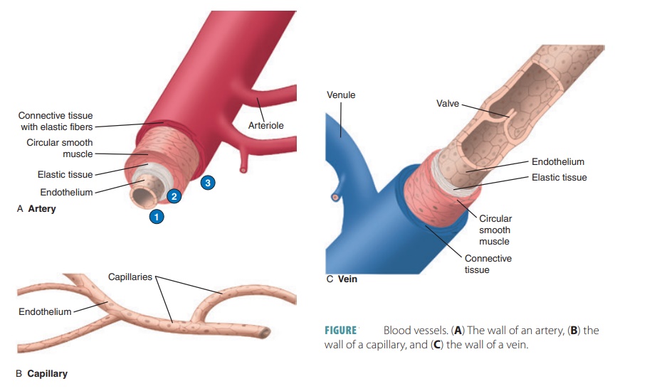
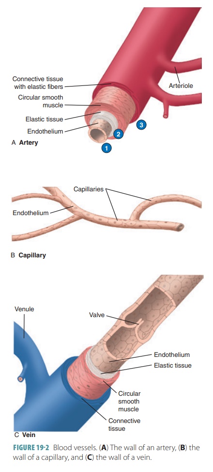
An artery’s wall consists of three distinct layers and blood-containing space known as the lumen.
Blood
Vessel Structure
An artery’s wall consists of three distinct layers and
blood-containing space known as the lumen. The
innermost tunica intima is made
up of a layer of simple squamous epithelium known as the endothelium (1). It rests on a
connective tissuemembrane with many elastic, collagenous fibers. The
endothelium helps prevent blood clotting and may also help in regulating blood flow. It releases nitric
oxide to relax smooth muscle of the vessel. In arter-ies, the outer margin has
a thick layer of elastic fibers known as the internal elastic membrane.
The middle tunica
media (2) makes up most of an arterial wall, including smooth
muscle fibers arranged mostly in circles and a thick elastic con-nective tissue
layer. The smooth muscle fiber activity is controlled by many chemicals and the
autonomic nervous system’s sympathetic vasomotor
nerve fibers. The tunica media is separated from the next layer, the tunica
externa, by a thin band of fibers known as the external elastic membrane.
The outer tunica externa (3), also known as the tunica adventitia, is thinner, made mostly of connective tissue with irregular fibers. It is attached to the sur-rounding tissues (FIGURE 19-2) and contains many lymphatic vessels and nerve fibers. The tunica externa of larger blood vessels contains many tiny blood vessels, which comprise a system known as the vasavasorum. This system nourishes the outer tissuesof blood vessel walls. The luminal inner portions of vessels obtain nutrients directly from the blood.
In arteries and arterioles, vasomotor fibers receive impulses to contract and
reduce blood vessel diameter, a process known as vasoconstriction. When inhibited, the muscle fibers
relax and the vessel’s diameter increases in a process known as vasodilation. Changes in artery and arteriole
diame-ters greatly affect blood flow and pressure.
