Neural Controls of Blood Vessels
| Home | | Anatomy and Physiology | | Anatomy and Physiology Health Education (APHE) |Chapter: Anatomy and Physiology for Health Professionals: Vascular System
1. Describe baroreceptor reflexes that control blood pressure changes. 2. Explain the location of chemoreceptors and their roles. 3. Describe how ADH and aldosterone regulate blood pressure. 4. Explain short-term and long-term regulation of blood pressure.
Neural
Controls of Blood Vessels
Most neural controls of blood vessels operate because of
reflex arcs, which involve baroreceptors and related afferent fibers. The reflexes
are controlled by the car-diovascular center of the medulla in the brain. Their
output travels thorough autonomic fibers to the heart and vascular smooth
muscle. The neural control mechanism is sometimes influenced by input from
chemoreceptors and higher brain centers.
Sympathetic efferents known as vasomotor fibers are used to transmit highly steady impulses from
the vasomotor center, which controls
blood vessel diameter. Nerves arising from the vasomotor center of the medulla
oblongata change, in cycles, the diameter of the lumen of each blood vessel,
controlling the volume of blood that is contained. Vasomotor fibers emerge from
the T1 through the L2 levels of the spinal cord and innervate the smooth
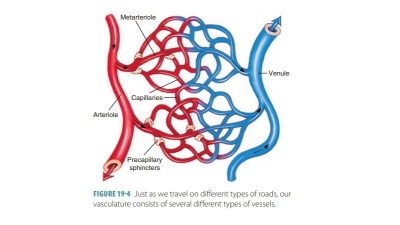
muscle of primarily arterioles but also of other blood vessels. This means the
arterioles are nearly always slightly contracted. This is known as vasomotor tone, which is different between various
body organs. For example, vasomotor impulses are more frequent in the skin and
digestive viscera arterioles but less frequent in the skeletal mus-cles. As a
result, they have more constriction than in the skeletal muscles. Generalized
vasoconstriction and increased blood pressure result from any increase in
sympathetic activity. Vascular muscle can relax slightly because of decreased
sympathetic activity. This allows blood pressure to reduce to basal levels.
There are three ways that cardiovascular center activity is modified:
■■From baroreceptors, which respond to
arterial pressure changes and stretching
■■From chemoreceptors, which respond
to changes in carbon dioxide, hydrogen, and oxygen levels inthe blood
■■From the higher brain centers
Baroreceptor Reflexes
Baroreceptors
are activated by increased arterialblood pressure and are located
in the carotid sinuses,
which provide the brain’s major blood supply; in the aortic arch; and in the
walls of most large neck and thoracic arteries. Stretching causes the
baroreceptors to send impulses quickly to the cardiovascular center. This
inhibits the cardioacceleratory and vasomotor centers while stimulating the
cardioinhibitor center. Blood pressure decreases as a result of these actions.
Baroreceptors are also found in the aortic
sinuses of the ascending aorta of
the heart and wall of the right atrium. Atrial
baroreceptors monitor blood pressure at the vena cava and right atrium, which constitute the end of the
systemic circuit. The atrial
reflex responds to stretching of the wall of the right
atrium.
The circulation is buffered from acute changes in blood
pressure by the quick responses of the barore-ceptors. Primarily in the head,
blood pressure falls as we stand up after lying down. The blood supply to the
brain is protected by the baroreceptors and their actions in the carotid sinus reflex. The baro-receptors that are
activated in the aortic
reflex help balance blood pressure in the overall systemic
circuit. Sustained pressure changes, such as chronic hyper-tension, usually
override the effects of baroreceptors. The baroreceptors become adapted to
monitor pres-sure changes at the new, higher “set point.”
Chemoreceptor Reflexes
Chemoreceptors
in theaortic archand
carotid arter-ies send impulses to the cardioacceleratory center, increasing
cardiac output. They also send impulses to the vasomotor center, causing reflex
vasoconstriction. Chemoreceptors act with chemoreceptor reflexes when carbon dioxide
levels rise, pH falls, or blood oxygen levels drop quickly. The resultant blood
pres-sure increase causes blood to return to the heart and lungs more quickly.
The carotid and aortic bodies close to the baroreceptors of the carotid sinuses and
aortic arch are the most important chemoreceptors. They play a greater role in
regulating respiratory rate, how-ever, than blood pressure.
High Brain Center Influences
The brain stem’s medulla oblongata integrates reflexes that
maintain blood pressure. The cerebral cortex and hypothalamus have the ability
to change arterial pressure by using relays to the centers of the medulla
oblongata. The fight-or-flight response is an example. It is controlled by the
hypothalamus, with large effects on blood pressure. Redistribution of blood
flow and other cardiovascular responses is also regulated by the hypothalamus.
Examples of this redistribution include during body temperature changes and exercise.
Short-Term Regulation by Hormonal Controls
Hormonal controls help to control blood pressure in
short-term peripheral resistance changes as well as long-term blood volume
changes. Local chemicals known as paracrines
help to bring adequate blood flow to servecertain tissues’
metabolic needs. Rarely, large releases of paracrines can affect blood
pressure. Short-term hor-monal controls involve antidiuretic hormone (ADH),
angiotensin II, atrial natriuretic peptide (ANP), eryth-ropoietin, and the
hormones of the adrenal medulla.
Antidiuretic Hormone
Antidiuretic
hormone (ADH) is also calledvasopressin.It is
produced by the hypothalamus, and released from the posterior lobe of the
pituitary gland, due to a decrease in blood volume. It may also be caused by an
increase in plasma osmotic concentration, or a secondary increase in
circulating angiotensin II. Antidiuretic hormone stimulates the kidneys to
conserve water. Although not usually important for regulation of blood pressure
on a short-term basis, if blood pressure falls to extremely low levels, its
release is greatly increased. Severe hemorrhage is an example of a situation
that triggers this release. ADH then helps to restore arterial blood pressure
via extensive peripheral vasoconstriction.
Angiotensin II
Angiotensin
II is generated within the specialized jux-taglomerular cells of the kidneys, by the enzymaticactions of renin. The kidneys release renin when
blood pressure or volume is low.
The steps of the effects of renin are as follows:
■■ Renin converts the liver-produced
plasma protein called angiotensinogen
to angiotensin I.
■■ In the lung capillaries, angiotensin-converting enzyme (ACE) modifies
angiotensin I to angiotensin II, which is an active hormone that has many
effects.
Angiotensin
II has four important functions, as follows:
■■
It stimulates adrenal production of aldosterone.This causes sodium
retention and potassium loss by the kidneys.
■■
It stimulates secretion of ADH. This then stimulates water
reabsorption by the kidneys and complements the effects of aldosterone.
■■
It stimulates thirst. This results in increased fluid consumption.
The presence of ADH and aldosterone means that the additional consumed water is
retained, and blood volume is elevated.
■■
It stimulates cardiac output and causes arteriole constriction.
This elevates systemic blood pressure. Angiotensin II has four to eight times
the effect on blood pressure than norepinephrine.
Atrial Natriuretic Peptide
Atrial natriuretic peptide (ANP) is produced by the right
atrium of the heart. It helps to reduce blood pressure and volume. ANP is
produced in response to excessive stretching during diastole. It acts by
antago-nizing aldosterone, causing the kidneys to excrete more water and
sodium. This reduces blood volume and also results in generalized vasodilation.
Ventricular muscle cells, exposed to similar stimuli, produce a related
hor-mone known as brain natriuretic
peptide (BNP). Both ANP and BNP reduce blood volume and pressure. They
accomplish this task in the following ways:
■■ By increasing sodium ion excretion from the kidneys
■■ By promoting water loss from increasing the volume of
urine produced
■■ By reducing thirst
■■ By blocking release of ADH, aldosterone,epinephrine, and
norepinephrine
■■ By stimulating peripheral vasodilation
When blood volume and pressure decline, the stress on the
heart walls is removed. Therefore, production of natriuretic peptide stops.
Adrenal Medulla Hormones
Adrenal
medulla hormones include epinephrine andnorepinephrine, which are released by
the adrenal gland during times of stress. In the blood these hor-mones increase
cardiac output and promote generalized vasoconstriction, enhancing the
sympathetic response. Generally, sympathetic stimulation, which releases
epinephrine, causes vasoconstriction and therefore increased blood pressure.
However, the parasympa-thetic nervous system has the opposite effect and
gen-erally causes vasodilation and decreased blood pressure.
Erythropoietin
■■ It stimulates adrenal production of
aldosterone. This causes sodium retention and potassium loss by the
kidneys.when blood pressure falls, or when the blood’s oxygen content becomes
abnormally low. It acts directly on the blood vessels. Vasoconstriction
increases blood pressure. The production and matura-tion of red blood cells is
also stimulated by EPO. These cells increase blood volume and viscosity. They
also improve its capacity to carry oxygen.
Long-Term Regulation by Renal Controls
Long-term control of blood pressure involves the kid-neys.
This alters blood volume instead of peripheral resistance and cardiac output.
There are two mecha-nisms: direct and indirect.
Direct Renal Mechanism
The direct renal mechanism changes blood volume without
using hormones. The rate of fluid filtering from the bloodstream to the kidney
tubules becomes faster when either blood pressure or blood volume rises. When
this occurs, more fluid leaves the body in urine because the kidneys cannot
reabsorb the fil-trate quickly enough. Therefore, both blood pressure and
volume are lowered. When they are low, water is conserved. It is returned to
the bloodstream, and the blood pressure increases.
Indirect Renal Mechanism
The indirect renal mechanism uses the renin-
angiotensin-aldosterone mechanism. Renin
is an enzyme that is released by certain kidney cells into the blood when
arterial blood pressure declines. It causes enzymatic claving of angiotensinogen, which is a plasma
protein manufactured by the liver. Renin converts angiotensinogen to angiotensin I. Then, angiotensin-converting enzyme converts angiotensin
Ito angiotensin II. The activity of
angiotensin-converting enzyme is linked with the capillary endothelium
pri-marily in the lungs but also in other body tissues.
There are four ways in which angiotensin II sta-bilizes
extracellular fluid volume and arterial blood pressure:
■■ Angiotensin II stimulates the
adrenal cortex to secrete aldosterone. This
hormone enhances renal absorption of sodium. Sodium moves into the bloodstream,
followed by water, conserving blood volume. Angiotensin II also directly
stimu-lates the kidneys’ reabsorption of sodium.
■■ Angiotensin II causes the posterior
pituitary to release ADH. This promotes additional water reabsorption by the
kidneys.
■■ Angiotensin II increases the thirst
sensation via activation of the hypothalamic thirst center. Water consumption
therefore increases, restoring blood volume and blood pressure.
■■ Angiotensin II is a very potent
vasoconstrictor. It increases peripheral resistance, which increases blood
pressure.
Homeostatic Imbalances
Homeostatic imbalances in blood pressure involve
hypertension and hypotension. Hypertension is
chronically elevated blood pressure, defined as a sus-tained increase in either
systolic pressure or diastolic pressure. In hypertension, systolic pressure is
usually above 140 mm Hg and diastolic pressure is usually above 90 mm Hg.
Chronic hypertension is common and dangerous because the heart must pump harder
against greater resistance, causing the myocardium to enlarge. Nearly 90% of
hypertensive patients have primary or essential hypertension, which has no
iden-tified, underlying cause. Primary hypertension may be linked to heredity,
diet, obesity, age, diabetes mellitus, stress, and smoking. It can usually be
con-trolled but cannot be cured. Secondary
hypertension is from an identifiable condition such as kidney dis-ease,
renal artery obstruction, hyperthyroidism, or Cushing’s syndrome.
Hypotension,
defined as blood pressure below90/60 mm Hg, is often linked simply to old age.
It is usually only dangerous if it leads to dizziness or faint-ing and, when
acute, is an important sign of circu-latory shock. Orthostatic hypotension is a temporary blood pressure drop, which
causes dizziness, when a person stands up suddenly after sitting or lying down.
It is most common in the elderly. Chronic
hypotension may be linked to a more serious disorder such as Addi-son’s
disease, hypothyroidism, or severe malnutrition.
1. Describe baroreceptor reflexes that control blood pressure
changes.
2. Explain the location of chemoreceptors and their roles.
3. Describe how ADH and aldosterone regulate blood pressure.
4. Explain short-term and long-term regulation of blood
pressure.