Factors of Respiratory Rate and Depth
| Home | | Anatomy and Physiology | | Anatomy and Physiology Health Education (APHE) |Chapter: Anatomy and Physiology for Health Professionals: Respiratory System
The depth of inspiration during breathing is based on the level of activity of the respiratory center and its stimulation of motor neurons that serve the respiratory muscles.
Factors of Respiratory Rate and Depth
The depth of inspiration during breathing is based on the level of activity of the respiratory center and its stimulation of motor neurons that serve the respiratory muscles. With more stimulation, increased num-bers of motor units are excited. Therefore, respiratory muscles contract with greater force. Respiratory rate is established by the length of time the inspiratory center is active or how fast it is turned off. Deep breathing is referred to as diaphragmatic breathing, while shallow breathing is known as costal breathing.
Certain chemicals also affect respiratory rate and depth. Important substances include CO2, hydrogen, and oxygen ions in the arterial blood. Other factors include emotional states, lung stretching capability, and levels of physical activity. Chemosensitive areas known as central chemoreceptors, located in the medulla oblongata, sense CO2 and hydrogen ion changes in the cerebrospinal fluid. When these levels change, respira-tory rate and TV are signaled to increase. More CO2 is exhaled, and both blood and cerebrospinal fluid levels of these chemicals fall, decreasing breathing rate.
CO2 is the most important chemical regulator of respiration. Arterial partial pressure of CO2 is usually 40 mm Hg, maintained within 3 mm Hg of this level, mostly by how rising CO2 levels affect the central che-moreceptors. Hypercapnia is a condition in which CO2 accumulates in the brain. The accumulating CO2 is hydrated and H2CO3 is formed. When the acid is dissociated, hydrogen ions are freed and pH drops. This also happens when CO2 enters red blood cells.
Increased hydrogen ions excite the central chemore-ceptors, which extensively synapse with the respiratory regulatory centers. Breathing depth and rate, therefore, increase. Because alveolar ventilation is enhanced, CO2 is quickly flushed out of the blood and pH rises. Alveolar ventilation is doubled with an elevation of only 5 mm Hg in arterial partial pressure of CO2. This is true even when there is no change in arterial oxygen levels or pH. The response to elevated partial pressure of CO2 is even more extensive when partial pressure of oxygen and pH are lower than normal. Increased ventilation is usually self-limited. It stops when there is restoration of homeostatic blood partial pressure of CO2.
The rising levels of hydrogen ions within the brain increase the activity of the central chemoreceptors, even though rising blood CO2 is the first stimulus. Although hydrogen does not easily diffuse across the blood–brain barrier, CO2 accomplishes this with no problem. Therefore, control of breathing while resting mostly is based on regulation of hydrogen ion concen-tration in the brain.
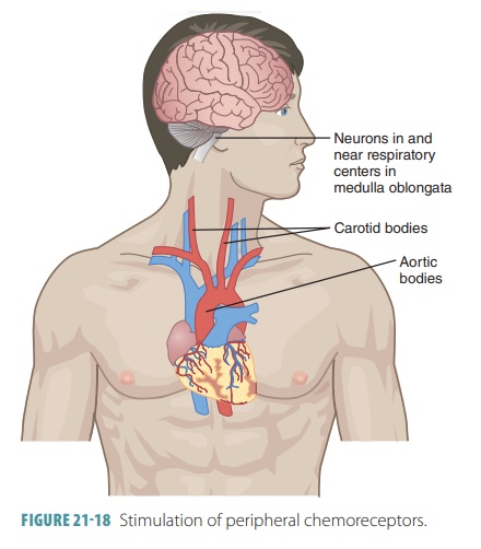
However, peripheral chemoreceptors in the carotid and aortic bodies also help and are able to sense changes in blood oxygen levels (FIGURE 21-18). Then, they increase the breathing rate, but this action requires extremely low levels of blood oxygen to occur.
The depth of breathing is regulated by the inflation reflex, which occurs when stretched lung tissues stimu-late stretch receptors in the visceral pleura, bronchioles, and alveoli. The duration of inspiratory movements is shortened, preventing overinflation of the lungs during forceful breathing. Emotional upset such as that caused by fear and pain usually increase breathing rate. If breathing stops, even for a short time, blood levels of CO2 and hydrogen ions rise and oxygen levels fall. Chemoreceptors are stimulated and the urge to inhale increases, overcoming the lack of oxygen. The deflation reflex usually only functions during forced exhalation and inhibits the expiratory centers while stimulating the inspiratory centers when the lungs are deflating.
How Partial Pressure of Oxygen Influences Breathing
The peripheral chemoreceptors contain cells that are sensitive to arterial levels of oxygen. These chemo-receptors lie in the aortic bodies of the aortic arch and the carotid bodies at the bifurcation of the com-mon carotid arteries. Those in the carotid bodies are the main oxygen sensors. Normally, reducing par-tial pressure of oxygen only affects ventilation mini-mally. This primarily involves enhanced sensitivity of peripheral receptors to increased partial pressure of CO2. For oxygen levels to become a strong stimulus for increased ventilation, arterial partial pressure of oxygen must drop greatly, to at least 60 mm Hg.
How Arterial pH Influences Breathing
Even during normal levels of oxygen and CO2, changes to arterial pH can modify the rate and rhythm of breathing. Increased ventilation occurring because of reduced arterial pH is controlled via the peripheral chemoreceptors. This is in part because hydrogen ions do not cross the blood–brain barrier. Changes in par-tial pressure of CO2 and hydrogen ion concentration are related, yet different.
Reduced blood pH may be related to retention of CO2. However, it may also occur because of meta-bolic reasons. These include lactic acid accumulation because of exercise or fatty acid metabolite or ketone body accumulation because of uncontrolled diabetes mellitus. No matter what the reason, as arterial pH declines, the respiratory system will attempt to com-pensate and raise the pH. This occurs by the increase of respiratory rate and depth in an attempt to elimi-nate CO2 and H2CO3 from the blood.
How Higher Brain Centers Influence Breathing
Respiratory rate and depth are modified when pain or strong emotions send signals to the respiratory centers. This occurs via the limbic system, including the hypothalamus. Changes in body temperature also affect respiration, with hotter temperatures increasing it and colder temperatures decreasing it.
Conscious control of breathing can also occur. The cerebral motor cortex sends impulses to the motor neurons, causing stimulation of respiratory muscles. This bypasses the medullary centers. Holding the breath is a limited function because the brain stem respiratory centers automatically reinitiate breathing once CO2 levels in the blood become critical.