Gross Anatomy of the Lungs
| Home | | Anatomy and Physiology | | Anatomy and Physiology Health Education (APHE) |Chapter: Anatomy and Physiology for Health Professionals: Respiratory System
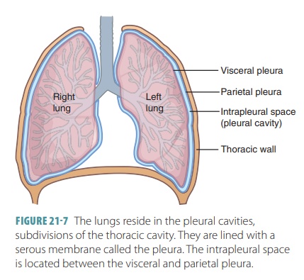
The paired lungs are located in the thoracic cavity and consist of soft, spongy, cone-shaped tissue. The right and left lungs are separated medially by the mediastinum and are enclosed by the thoracic cage and diaphragm.
Organization of the Respiratory System
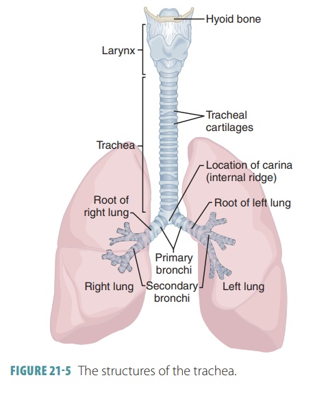
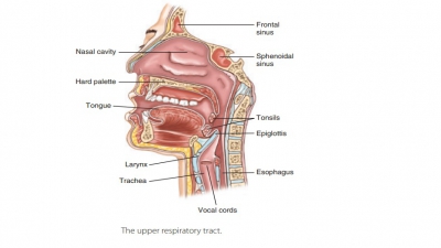
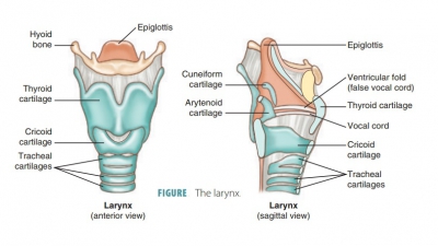
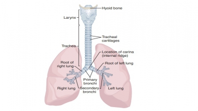
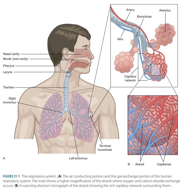
The upper respiratory tract includes the nose, nasal cavity, paranasal sinuses, and pharynx. The lowerrespiratory tract includes the larynx, trachea, and lungs. The lungs contain the bronchi, bronchioles, and alveoli. FIGURE 21-1 shows the structures of the respiratory system.
Gross
Anatomy of the Lungs
The paired lungs are
located in the thoracic cavity and consist of soft, spongy, cone-shaped tissue.
The right and left lungs are separated medially by the mediastinum and are
enclosed by the thoracic cage and diaphragm. The
mediastinum contains the heart, major blood vessels, bronchi, esophagus, and
various organs. Air is inhaled by the active contraction of the respiratory
muscles, including the diaphragm. Each lung occupies most of the available
thoracic space on its side and is suspended by a bronchus and large blood
vessels that enter on the lung’s medial surface. The collective term for each
lung’s vascular and bronchial attachments is the root. Just below the clavicle bone on each site is the lung’s
narrow superior tip or apex. The base of the lung is the concave inferior
surface that rests on the dia-phragm muscle. Each lung’s mediastinal surface
has an indentation known as the hilum.
Pulmonary and systemic blood vessels and bronchi, lymphatic vessels, and nerves
enter and leave the lung at this point.
The lungs are not symmetrical. The three-lobed right lung is larger because the two- lobed left lung’s space has to accommodate the heart as well. The left lung has a cardiac notch, which is a concavity in its medial aspect that accommodates the heart. Each lung lobe is supplied by a major branch of the bron-chial tree and is connected to blood and lymphatic vessels. In the right lung, the superior and middle lobes are separated by the horizontal fissure and the middle and inferior lobes are separated by an inferior oblique fissure. In the left lung, the fissure separat-ing the superior and inferior lobes is also called the oblique fissure.
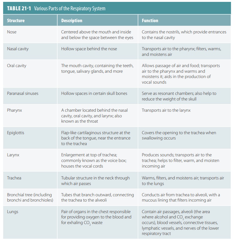
Each lung contains air passages, alveoli, blood vessels,
connective tissues, lymphatic vessels, and nerves. TABLE 21 -1 lists the parts of the respiratory system.
Atthe lung surface, resembling hexagons are the lung lob-ules, the smallest subdivisions of the lungs that can beseen
without a microscope, which are less than 1 inch in size. Each lobule is served
by a large bronchiole and its branches. Smoking or regularly inhaling pollution
causes the connective tissue between the lobules to turn black as they collect
carbon.
While most of the lungs contain air spaces, the remainder is
made up of the stroma, which consists
primarily of elastic connective tissue. This makes the lungs very soft, spongy,
and light. They collectively weigh slightly more than 1 kg or 2.2 pounds. Their
elasticity makes the ability to breathe easier. The elastic components of the
lungs recoil when the mus-cles of inhalation relax. The action of the diaphragm
and rib cage returning to their normal positions is called elastic rebound.