Larynx
| Home | | Anatomy and Physiology | | Anatomy and Physiology Health Education (APHE) |Chapter: Anatomy and Physiology for Health Professionals: Respiratory System
1. Identify the structures of the upper respiratory system. 2. What are the functions of the paranasal sinuses? 3. Name the laryngeal cartilages. 4. Discuss how the vocal cords produce the sounds used in speech.
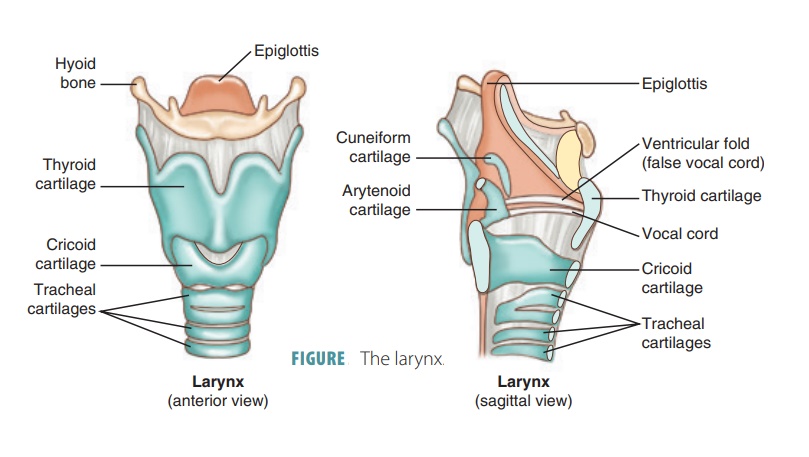
Organization of the Respiratory System
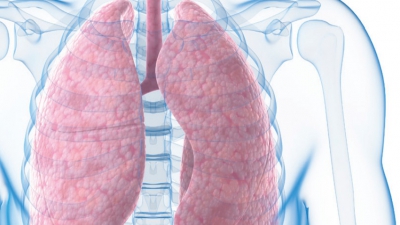
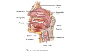
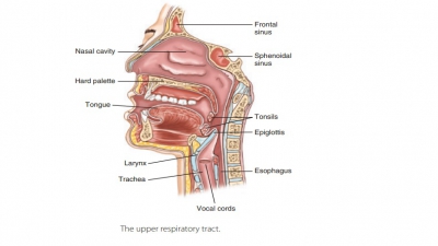
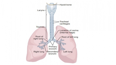
The upper respiratory tract includes the nose, nasal cavity, paranasal sinuses, and pharynx. The lowerrespiratory tract includes the larynx, trachea, and lungs. The lungs contain the bronchi, bronchioles, and alveoli. FIGURE 21-1 shows the structures of the respiratory system.
Larynx
An enlargement in the airway above the trachea and below the
pharynx is called the larynx,
com-monly called the voice box. The
larynx is about 5 cm or 2 inches in length, extending from the level of the
third to the sixth cervical vertebra. It attaches to the hyoid bone superiorly,
opening into the laryngophar-ynx, and is continuous with the trachea
inferiorly.
The larynx controls how air and food are passed into their
proper channels. It also functions to produce a person’s voice. It conducts air
into and out of the tra-chea while preventing foreign objects from entering,
and houses the vocal chords. The larynx is made up of muscles and nine
cartilages that are bound by elas-tic tissues, consisting of ligaments and
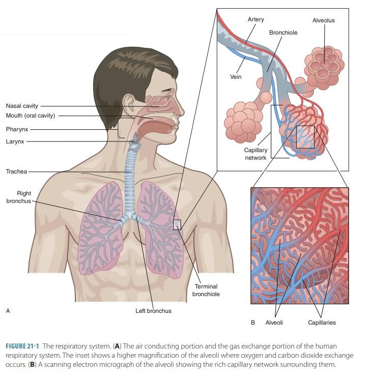
membranes. The cartilages include the thyroid cartilage, cricoidcartilage, and epiglottic cartilage (FIGURE 21-3). Allthe laryngeal
cartilages, except for the epiglottis, are hyaline
cartilages. These cartilages are bound to eachother by intrinsic ligaments.
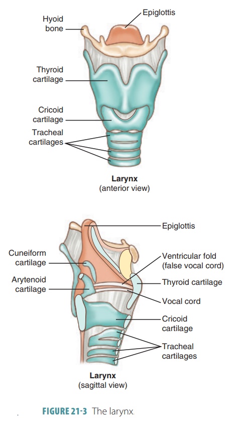
Two cartilage plates fuse to form the large thy-roid cartilage, which resembles a
shield in shape. Atits midline, a laryngeal
prominence exists, which is externally visible. This is commonly known
as the Adam’s apple. In males, this
is usually larger and itsgrowth is stimulated by male sex hormones during
puberty. The thyroid cartilage makes up most of the anterior and lateral
surface of the larynx. The ring-shaped cricoid
cartilage is inferior to the thyroid cartilage and is located above the
trachea, to which it is inferiorly anchored. Parts of the lateral and
poste-rior laryngeal walls are formed by three pairs of car-tilages. These are
called the arytenoid cartilages, cuneiform
cartilages, andcorniculate
cartilages.The pyramid-shaped arytenoid cartilages are most important,
because they anchor the vocal folds. The arytenoid cartilage articulates with
the superior border of the cricoid cartilage.
The flap-like structure that actually allows the larynx to
“control” whether air or food passes is the epiglottis. It is actually the ninth cartilage of the
lar-ynx and extends from the tongue’s posterior aspect to where it is anchored,
on the anterior rim of the thy-roid cartilage. When swallowing, the larynx
rises and the epiglottis presses downward, partially covering the opening into
the larynx to help prevent foods and liq-uids from entering the air passages.
The epiglottis is spoon-shaped, highly elastic, and nearly covered by a mucosa
that contains taste buds.
During breathing, the larynx is wide open and the free edge
of the epiglottis projects upward. Anything besides air that enters the larynx
triggers the cough reflex so the substance can be expelled. However, the cough
reflex does not work when a person is uncon-scious. Liquids should, therefore,
never be given to an unconscious person.
Under the laryngeal mucosa, on each side, are the highly
elastic vocal ligaments that
attach the arytenoid cartilages to the thyroid cartilage. They form
horizon-tal vocal folds inside the larynx that extend inward and are divided
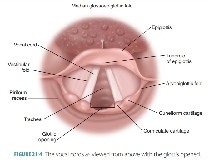
into upper and lower folds. The upper folds are called false vocal cords or vestibular
folds because they do not create sounds (FIGURE 21 -4); they help close the airway during
swallowing. The lower vocal folds are called true vocal cords because they actually create sounds when air is
forced between them, causing them to vibrate from side to side. In appearance,
the true vocal cords are pearly white in color because they lack blood vessels.
Using the tongue and lips to change the shape of the pharynx and oral cavity
transforms sound waves into words. The contraction or relaxation of the vocal
cords alters their tension, controlling the pitch they emit. Increasing tension
raises the pitch, whereas decreasing tension lowers the pitch. The loudness of
a sound is controlled by the force of air passing through the vocal cords.
During breathing, the glottis is a triangular
slit between the vocal cords. When food or liquid is swallowed, the glottis
closes to prevent it from entering the trachea.
The superior portion of the larynx is lined with stratified squamous epithelium, and comes into contact with food. Under the vocal folds, there is a pseudostratified ciliated columnar epithelium, which filters dust. Its cilia stroke upward, toward the phar-ynx. This continually moves mucus away from the lungs. When a person “clears the throat”, the action assists mucus in moving up and out of the larynx.
Sound Production
Sound waves are produced as air passes through the glottis
and vibrates the vocal folds. The pitch
of the sound is based on the vocal folds’ tension, diameter, and length. Their
diameter and length are closely related to the overall size of the larynx.
Tension is con-trolled by intrinsic laryngeal muscles, which cause dif-ferent
positions of the arytenoid cartilages, related to the thyroid cartilage. If
distance increases, the tension in the vocal folds increases, and the pitch
rises. Oppo-sitely, when distance decreases, the vocal folds relax and the
pitch lowers. In children, the vocal folds are thin and short, resulting in
higher pitched voices. The larynx increases in size during puberty in both
sexes.However, males experience a significantly greater increase, causing their
adult voices to become lower than those of adult females.
At the larynx, sound production is known as pho-nation, which is a part of speech
production. To speakclearly, articulation
is also required, which is the mod-ification of sounds that are created via the
tongue, lips, and teeth. Like a musical instrument, sound amplifica-tion and
resonance occur inside the pharynx, oral and nasal cavities, and paranasal
sinuses. These structures help to make each person’s voice distinct. Sound
pro-duction changes when the nasal cavity and paranasal sinuses become filled
with mucus instead of air, such as when sinus infections develop.
To produce distinct words, voluntary tongue, lip, and cheek
movements are used. When the lar-ynx becomes infected or inflamed (laryngitis), the vibrational qualities
of the vocal folds are usually affected. The voice usually sounds “hoarse” as a
result. If mild, this condition is temporary and not serious in most cases.
Bacterial or viral infections of the epiglottis can become very serious,
however. If swelling closes the glottis, it is possible for the indi-vidual to
suffocate. Thisacute epiglottitis can
quickly develop after a bacterial throat infection, with young children most
often affected.
1. Identify the structures of the upper respiratory system.
2. What are the functions of the paranasal sinuses?
3. Name the laryngeal cartilages.
4. Discuss how the vocal cords produce the sounds used in
speech.
Related Topics
