Nose and Paranasal Sinuses
| Home | | Anatomy and Physiology | | Anatomy and Physiology Health Education (APHE) |Chapter: Anatomy and Physiology for Health Professionals: Respiratory System
The only externally visible part of the respiratory system is the nose, which is the primary passageway for incoming air.
Organization of the Respiratory System
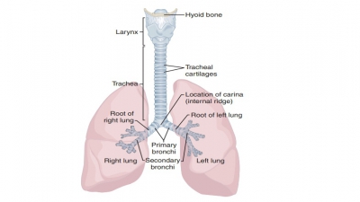
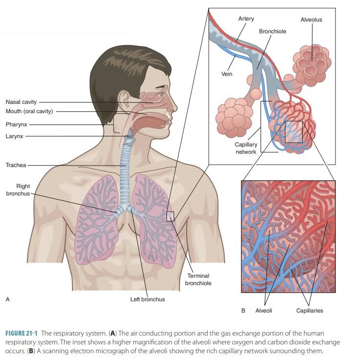
The upper respiratory tract includes the nose, nasal cavity, paranasal sinuses, and pharynx. The lowerrespiratory tract includes the larynx, trachea, and lungs. The lungs contain the bronchi, bronchioles, and alveoli. FIGURE 21-1 shows the structures of the respiratory system.
Nose and
Paranasal Sinuses
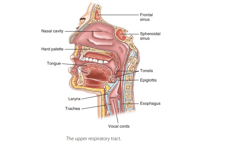
The only externally visible part of the respiratory system is the nose, which is the primary passageway for incoming air. It has a variety of respiratory func-tions, conditioning incoming air, filtering and clean-ing the air, functioning as a resonating chamber for speaking, and containing the smell or olfactory receptors. The conditioning of incoming air consists of moistening and warming.
The paired external
nares, commonly known as nostrils, open into the nasal cavity. The nasalvestibule is a space inside the
flexible nasal tissues.Its epithelium has coarse hairs extending across the
external nares. Large particles in the air, including dust, sand, and insects,
become trapped in these hairs, preventing them from entering the nasal cavity.
Nasal Cavity
The nasal
cavity is a hollow space located behind the nose divided into
right and left portions by the nasalseptum.
This structure is made up of the vomer boneand the perpendicular plate of the
ethmoid bone posteriorly and septal cartilage
anteriorly. Posteriorly, the nasal cavity is continuous with the nasal portion
of the pharynx via the posterior
nasal apertures, which are funnel-like structures also known as choanae.
The roof of the nasal cavity comprises the skull’s ethmoid
and sphenoid bones. Its floor is made up of the palate, a structure that separates it from the oral cavity below.
Anteriorly, the palate is supported by processes of the maxillary bones and the
palatine bones. This area is called the hard palate. There is also a soft palate, which is the unsupported poste-rior section.
Most of the nasal cavity is lined with respiratorymucosa, which is
pseudostratified ciliated epitheliumcontaining many scattered, mucus-secreting gobletcells. This epithelium
includes a network of bloodvessels and rests on a lamina propria, which has a
rich supply of seromucous nasal glands.
Every day, these glands secrete approximately 1 liter of mucus, which contains
the antibacterial enzyme known as lysozyme.
The filtration mechanisms that prevent contamination of the respiratory system
are referred to as the respira-tory
defense system.
As air passes through from the nostrils and over the mucosal
surfaces, heat from the blood in these underlying capillary plexuses warms the
air to more closely match the body’s temperature. Incoming air is also
moistened from H2O evaporation out of the mucous lining. The
lysozyme-containing mucus, pushed by the cilia of the epithelial lining,
entraps dust and other small particles and carries them toward the pharynx. The
lysozyme attacks and chemically destroys bacteria. The respiratory mucosa cells
also secrete defensins, which
function as natural antibiot-ics. The defensins kill invading microorganisms.
The respiratory mucosa has ciliated cells that create a slight current, moving
the contaminated mucus posteriorly toward the throat. Once swallowed, the mucus
and its contained microorganisms are destroyed by the gastric juice of the
stomach. The nasal cavity acts as a resonating chamber in speech.
There is a rich supply of sensory nerve endings in the nasal
mucosa. When they contact irritating dust, pollen, and other particles, the sneeze reflex is triggered. When we
sneeze, air is forced outward vio-lently, which effectively expels irritants.
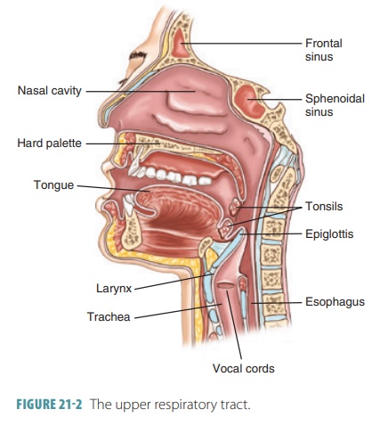
The bones and bone processes that project in a curved shape
from the nasal cavity’s lateral walls, dividing it into passages, are called
the nasal conchae (FIGURE 21-2). These include the superior, middle, and inferior nasal conchae. Together, they
increase themucosal surface area that is exposed to air as well as the air
turbulence in the nasal cavity.
Paranasal Sinuses
The paranasal
sinuses are a ring of air-filled spaces inside the skull bones that
open into the nasal cavity. Located inside the maxillary, frontal, ethmoid, and
sphenoid bones of the skull, they are lined with mucous membranes that are
continuous with the lining of the nasal cavity. These sinuses reduce the
skull’s weight and resonate, affecting the quality of the voice. They also help
to warm and moisten the incoming air. Mucus produced by the paranasal sinuses
eventually flows to the nasal cavity. When you blow your nose, a suction-ing
effect is created that helps to drain the sinuses.