Structure of Endomembrane System and Cytoskeleton
| Home | | Anatomy and Physiology | | Anatomy and Physiology Health Education (APHE) |Chapter: Anatomy and Physiology for Health Professionals: Levels of Organization : Cells
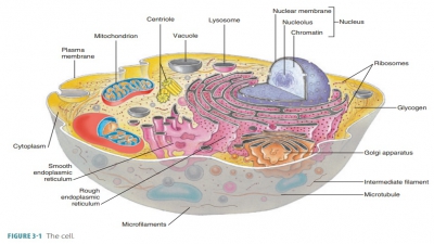
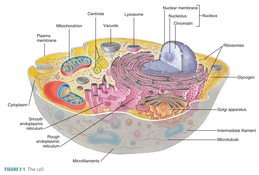
The endomembrane system consists of organelles that collectively produce and degrade biologic molecules (also storing and exporting them) and that degrade substances that may be harmful.
Endomembrane
System and Cytoskeleton
Endomembrane System
The endomembrane system consists of organelles that collectively
produce and degrade biologic molecules (also storing and exporting them) and
that degrade substances that may be harmful. This system is made up of the ER,
Golgi apparatus, secretory vesicles, lysosome, and nuclear membrane. Its
components include all membranous organelles or elements that are structurally
continuous or that arise due to fusing or forming transport vehicles.
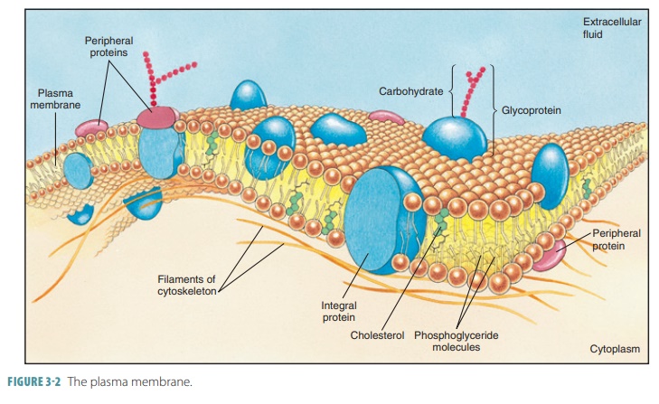
Continuities exist between the nuclear envelope, the RER, and the SER. Although
not an actual endomembrane, the
plasma membrane is also part of the endomembrane system. Throughout this
system, many indirect interactions occur. Certain vesicles that begin in the ER
are eventually fused with the Golgi apparatus or plasma membrane. Vesicles from
the Golgi apparatus can become part of the lyso-somes, plasma membrane, or
secretory vesicles.
Cytoskeleton
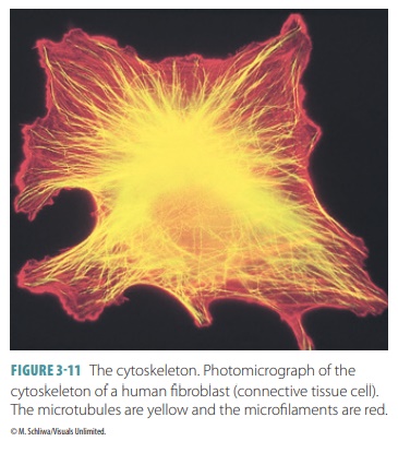
The cytoskeleton is a network of many rods and hun-dreds of proteins (FIGURE 3-11). The
rods run through the cytosol, whereas the proteins link them to other
structures of the cell. The cytoskeleton, therefore, functions as the cell’s
skeleton, muscles, and ligaments. Various cell movements are also generated by
the cyto-skeleton. The three types of rods in the cytoskeleton, all of which
lack covering membranes, are microfilaments,intermediate
filaments, and microtubules.
Microfilaments are the thinnest
type of cytoskel-etal rods, are semiflexible, and are made of actin, a protein. They are the most
fragile of the cytoskeletal elements. Because no two cells are identical, each
has its own unique microfilaments. However, almost all cells have a
cross-linked microfilament network (the terminal
web) that is attached to the cytoplasmic side of the plasma membrane. The
cell surface is strength-ened by this web, which also acts against compres-sion
and transmits force during shape changes and cellular movements.
Microfilaments are usually
involved in cell move-ment and shape changes. Actin filaments interact with unconventional myosin, another protein,
to generate contractile cellular forces. It is by this mechanism that cells are
“pinched” into two cells during cell division. Microfilaments are used for the
motion of amoeba and when membranes change during exocytosis and endocytosis.
In most cells except for muscle cells, actin filaments breakdown regularly and
reform from smaller subunits as needed.
Intermediate filaments resemble
ropes and are made of strong, insoluble protein fibers. Twisted tetramer fibrils form intermediate
filaments, which arethicker (in diameter) than microfilaments but thinner than
microtubules. Intermediate filaments have high tensile strength, and of all
cytoskeletal elements are the most permanent and stable. They attach to
desmo-somes to resist pulling forces that may be exerted on the cell. Their
protein composition differs in various cell types. Therefore, they are named
differently based on the cells they exist in. For example, in nerve cells, the
intermediate filaments are referred to as neurofilaments.
Microtubules are hollow
organelles, made up of spherical tubulins,
which are protein subunits. They usually radiate from a small area of cytoplasm
near the nucleus (the centrosome or cell center). Microtubules are always
active, growing from the centrosome, then dis-assembling, and then reassembling
in either different sites or the same site. They are stiff but bendable and
determine the cell’s overall shape as well as how cellu-lar organelles are
distributed. From the microtubules, structures appear to “hang.” These include
lysosomes, mitochondria, and secretory vesicles. Tiny motor pro-teinsconstantly move and reposition organelles along the
microtubules. These motor proteins include dyneins,kinesins,
and others. They function by changing shape,energized by ATP. Some move
substances along the microtubules evenly, whereas others grip and release the
microtubule, repeating these actions again and again.
Related Topics