Coagulants
| Home | | Medicinal Chemistry |Chapter: Medicinal Chemistry : Coagulants
Homeostasis is the cessation of blood loss from damaged vessels. Platelets first adhere to macromolecules in the subendothelial regions of injured blood vessels; they aggregate to form the primary haemostatic plug.
DRUGS ACTING ON BLOOD AND BLOOD FORMING ORGANS
Coagulants
INTRODUCTION
Homeostasis
is the cessation of blood loss from damaged vessels. Platelets first adhere to
macromolecules in the subendothelial regions of injured blood vessels; they
aggregate to form the primary haemostatic plug. Platelets stimulate the local
activation of plasma coagulation factors, leading to generation of a fibrin clot
that reinforces the platelet aggregate. Later, as wound healing occurs, the
platelet aggregates and fibrin clots are degraded. Thrombosis is a pathological
process in which a platelet aggregates and a fibrin clot occludes blood vessels.
Arterial thrombosis may result in ischaemic necrosis of tissues supplied by the
artery (e.g. myocardial infarction due to thrombosis of coronary artery).
Venous thrombosis may cause tissue drain by the vein to become edematous and
inflamed.
Coagulation
of blood comprises the formation of fibrin by a series of interactions among a
large number of protein factors and other substances. Blood coagulation process
requires coagulation factors, calcium, and phospholipids.
·The coagulation factors (proteins) are
manufactured by the liver.
·Ionized calcium (Ca++) is available
in the blood and from intracellular sources.
·Phospholipids are prominent components of the
cellular and platelet membranes. They provide a surface on which the chemical
reactions of coagulation can take place. The coagulation factors are numbered
in the order of their discovery.
Factor
I—Fibrinogen
Factor
II—Prothrombin
Factor
III—Tissue thromboplastin (tissue factor)
Factor
IV—Ionized calcium (Ca++)
Factor
V—Labile factor or proaccelerin
Factor
VI—Unassigned
Factor
VII—Stable factor or proconvertin
Factor VIII—Antihaemophilic
factor
Factor
IX—Plasma thromboplastin component, Christmas factor
Factor
X—Stuart–Prower factor
Factor XI—Plasma thromboplastin antecedent
Factor
XII—Hageman factor
Factor
XIII—Fibrin stabilizing factor
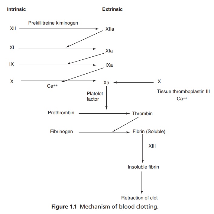
Mechanism of Blood Clotting
Coagulation
can be initiated by either of the two distinct pathways (Fig. 1.1):
1.
The
intrinsic pathway can be initiated by events that take place within the lumen
of blood vessels. This requires only elements (clotting factors, Ca++
platelet surface, etc) found within or intrinsic to the vascular system.
2.
The
extrinsic pathway is the other route to coagulation. It requires tissue factor
(tissue thromboplastin), a substance that is extrinsic to or not normally
cumulating in the vessel. Tissue factor is released when the vessel wall is
ruptured.
2. Calcium salts: Calcium salts, especially Ca++
intravenous injections, are very popular, but it does not help much unless
there is deficiency of Ca++ in the blood.
2 .Vitamin K (Synonym: Vitamin K1-Phytomenadione)
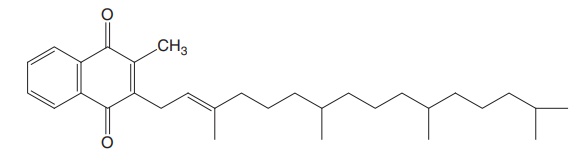
Properties and uses: Phytomenadione is a clear intense yellow viscous
oily liquid, practically insoluble in water, sparingly soluble in ethanol, and
miscible with fatty oils. Vitamin K is essential to keep up the prothrombin
level in blood by forming prothrombin in the liver. Hence, it is used orally
and intramuscular (IM), now water-soluble vitamin K is available for
intravenous (IV) administration. This is called methyl naphthaquinone and is
very useful in emergency.
Assay: It is assayed by adopting liquid chromatography technique.
Dosage forms: Phytomenadione injection B.P., Phytomenadione tablets B.P.
3. Vitamin K3 (Menadione)
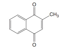
Properties and uses: Menadione is a pale-yellow crystalline powder,
practically insoluble in water, soluble in toluene, sparingly soluble in
alcohol and methanol. Used as source of vitamin K and has prothrombogenic
property.
Assay: Dissolve the sample in glacial acetic acid and add dilute
hydrochloric acid and zinc powder. Allow the mixture to stand and titrate
against 0.1 M ammonium cerric nitrate using ferroin as indicator.
Anticoagulants
Anticoagulants
are drugs that prevent the clotting of blood. Heparin is a glucosaminoglycan
found in the secretory granules of mast cells. It is synthesized from uridine
diphosphate sugar precursor as a polymer of alternating D-gluconic
acid and N-acetyl-D-glucosamine
residue. About 10–15 glucosaminoglycan chains, each containing 200–300
monosaccharide units, are attached to a core protein and yield a proteoglycan
with a molecular mass of 750,000–1,000,000 daltons. The glucosaminoglycan then
undergoes a series modification, which includes n-acetylation and n-sulphonation
of glucosamine, epimerization of D-gluconic acid to L-iduronic acid,
O-sulphation of iduronic and
glucoronic acid residues at the C2 position, and O-sulphation of glucosamine residue at C3 and C6
position. Each of these modification reactions is incomplete, yielding variety
of oligosaccharide structures. After the heparin proteoglycan has been
transported to the mast cell granule, an endo-β-D-glucuronidase
degrades the glycosamionoglycan chains to 5000–30,000 dalton fragments over a
period of hours.
Related Topics
